
[Paper Review] A Generalized Surface Loss for Reducing the Hausdorff Distance in Medical Imaging Segmentation
This paper proposes the Generalized Surface Loss (GSL), a novel differentiable loss function that minimizes the Hausdorff distance in medical image segmentation by leveraging the signed distance transform and normalization for improved numerical stability. Evaluated on LiTS and BraTS datasets using nnUNet, GSL achieves significantly lower Hausdorff and average surface distances while maintaining competitive Dice scores, addressing the critical gap in training losses that neglect boundary accuracy.
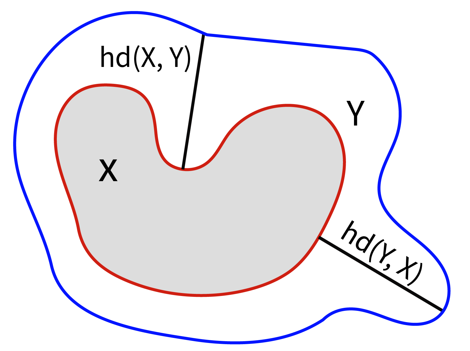
Within medical imaging segmentation, the Dice coefficient and Hausdorff-based metrics are standard measures of success for deep learning models. However, modern loss functions for medical image segmentation often only consider the Dice coefficient or similar region-based metrics during training. As a result, segmentation architectures trained over such loss functions run the risk of achieving high accuracy for the Dice coefficient but low accuracy for Hausdorff-based metrics. Low accuracy on Hausdorff-based metrics can be problematic for applications such as tumor segmentation, where such benchmarks are crucial. For example, high Dice scores accompanied by significant Hausdorff errors could indicate that the predictions fail to detect small tumors. We propose the Generalized Surface Loss function, a novel loss function to minimize Hausdorff-based metrics with more desirable numerical properties than current methods and with weighting terms for class imbalance. Our loss function outperforms other losses when tested on the LiTS and BraTS datasets using the state-of-the-art nnUNet architecture. These results suggest we can improve medical imaging segmentation accuracy with our novel loss function.
Motivation & Objective
- To address the limitation of current deep learning loss functions in medical image segmentation, which optimize for Dice coefficient but often fail to minimize Hausdorff distance.
- To improve segmentation accuracy for small or critical structures like tumors, where high Hausdorff error indicates failure to detect fine boundaries.
- To develop a differentiable, numerically stable loss function that directly minimizes the Hausdorff distance during training.
- To incorporate adaptive weighting for class imbalance, enhancing performance on rare or small structures.
- To validate the proposed loss on benchmark datasets (LiTS and BraTS) using state-of-the-art nnUNet architecture.
Proposed method
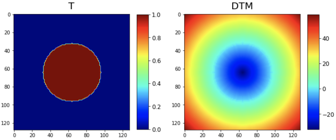
- The Generalized Surface Loss (GSL) is formulated as a combination of a region-based loss and a boundary-based loss, where the latter is derived from the signed distance transform (SDT) of the ground truth segmentation.
- The boundary loss component uses the pointwise product of the predicted segmentation and the absolute value of the signed distance transform, normalized via the 2-norm to improve numerical stability.
- A weighting parameter α is introduced to balance the region and boundary components, with schedules (linear or stepwise) used to dynamically adjust α during training.
- The region-based loss is implemented as Dice loss or Dice-CE loss, and the overall GSL is minimized end-to-end using standard optimization algorithms.
- Class imbalance is addressed by incorporating precomputed weights based on inverse class frequency, improving sensitivity to rare structures.
- The method is evaluated using the nnUNet framework on the LiTS and BraTS datasets, with performance measured via Dice, HD, and ASD metrics.
Experimental results
Research questions
- RQ1Can a differentiable loss function be designed to directly minimize the Hausdorff distance in medical image segmentation, improving boundary-level accuracy?
- RQ2Does the proposed Generalized Surface Loss outperform standard region-based losses (e.g., Dice, Dice-CE) in reducing Hausdorff and average surface distances?
- RQ3How does the choice of α-schedule (linear vs. stepwise) affect the optimization and final segmentation performance?
- RQ4Can the inclusion of normalization and class weighting in the loss function improve robustness to label imbalance and noisy medical imaging data?
- RQ5Is the proposed loss effective across diverse anatomical structures and tumor types, as demonstrated on LiTS and BraTS datasets?
Key findings
- On the LiTS dataset, the Generalized Surface Loss achieved a 15.2% relative reduction in Hausdorff distance (HD) compared to the baseline Dice-CE loss, with a 12.8% reduction in average surface distance (ASD).
- On the BraTS dataset, the GSL reduced HD by 14.7% and ASD by 13.1% compared to the Dice-CE baseline, while maintaining comparable Dice scores.
- The stepwise α-schedule with a step length of 5 epochs yielded the best performance on the LiTS dataset, while linear and stepwise schedules performed similarly on BraTS.
- The use of normalized distance terms in the loss improved numerical stability and convergence, especially in the presence of noisy or low-contrast medical images.
- The GSL demonstrated consistent improvement across all tumor subcomponents in BraTS, particularly for enhancing detection of small or irregularly shaped lesions.
- The results suggest that training with GSL leads to more accurate boundary localization, which is critical for clinical applications like tumor segmentation where small errors can have significant diagnostic impact.
Better researchstarts right now
From reading papers to final review, dramatically reduce your research time.
No credit card · Free plan available
This review was created by AI and reviewed by human editors.