
[Paper Review] Analysis of the BraTS 2023 Intracranial Meningioma Segmentation Challenge
This paper analyzes the BraTS 2023 Intracranial Meningioma Segmentation Challenge, evaluating deep learning models for segmenting meningiomas on pre-operative MRI. It identifies skull-stripping as a major issue causing loss of tumor volume, and recommends alternative anonymization techniques like MRI_reface to preserve critical tumor regions for radiotherapy planning.
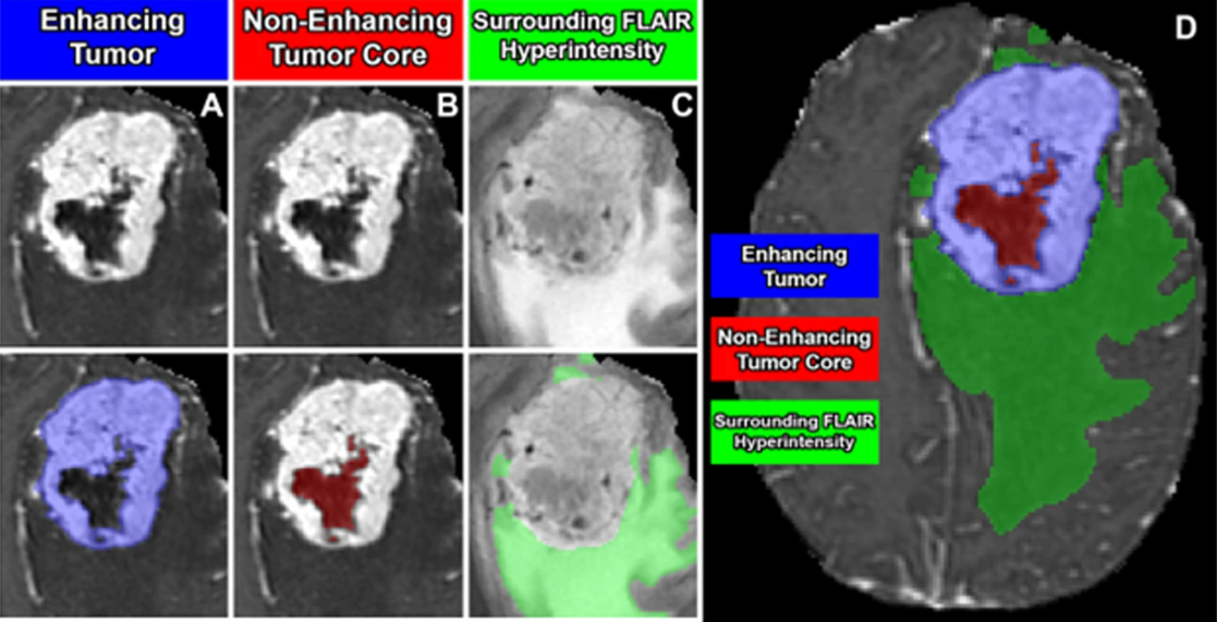
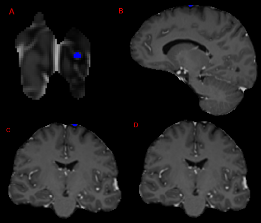
We describe the design and results from the BraTS 2023 Intracranial Meningioma Segmentation Challenge. The BraTS Meningioma Challenge differed from prior BraTS Glioma challenges in that it focused on meningiomas, which are typically benign extra-axial tumors with diverse radiologic and anatomical presentation and a propensity for multiplicity. Nine participating teams each developed deep-learning automated segmentation models using image data from the largest multi-institutional systematically expert annotated multilabel multi-sequence meningioma MRI dataset to date, which included 1000 training set cases, 141 validation set cases, and 283 hidden test set cases. Each case included T2, FLAIR, T1, and T1Gd brain MRI sequences with associated tumor compartment labels delineating enhancing tumor, non-enhancing tumor, and surrounding non-enhancing FLAIR hyperintensity. Participant automated segmentation models were evaluated and ranked based on a scoring system evaluating lesion-wise metrics including dice similarity coefficient (DSC) and 95% Hausdorff Distance. The top ranked team had a lesion-wise median dice similarity coefficient (DSC) of 0.976, 0.976, and 0.964 for enhancing tumor, tumor core, and whole tumor, respectively and a corresponding average DSC of 0.899, 0.904, and 0.871, respectively. These results serve as state-of-the-art benchmarks for future pre-operative meningioma automated segmentation algorithms. Additionally, we found that 1286 of 1424 cases (90.3%) had at least 1 compartment voxel abutting the edge of the skull-stripped image edge, which requires further investigation into optimal pre-processing face anonymization steps.
Motivation & Objective
- To evaluate the performance of deep learning models in segmenting intracranial meningiomas using the BraTS 2023 dataset.
- To investigate the impact of skull-stripping pre-processing on meningioma segmentation accuracy.
- To identify clinical relevance gaps in current segmentation tasks, particularly regarding tumor volume inclusion for radiation therapy planning.
- To recommend improved pre-processing techniques that preserve tumor volume while ensuring patient anonymization.
- To align automated segmentation with clinical protocols such as RTOG 0539 for target volume definition in meningioma radiotherapy.
Proposed method
- Utilized the BraTS 2023 pre-operative meningioma dataset comprising 1,424 cases with multi-contrast MRI (T1, T1-CE, T2, FLAIR).
- Evaluated segmentation performance using standard metrics on training (70%), validation (10%), and private test (20%) splits.
- Analyzed the effect of skull-stripping on tumor volume by measuring voxels abutting the skull boundary.
- Proposed alternative anonymization techniques such as MRI_reface, which modifies facial and ear regions to preserve brain tissue.
- Assessed the clinical relevance of segmentation outputs by comparing with RTOG 0539 guidelines for target volume definition.
- Evaluated the inclusion of non-enhancing compartments (e.g., T2/FLAIR hyperintensity, dural tails) in segmentation labels versus clinical practice.
Experimental results
Research questions
- RQ1To what extent does skull-stripping during pre-processing lead to the exclusion of meningioma tumor volume in automated segmentation?
- RQ2How do current segmentation models perform on the BraTS 2023 meningioma dataset, particularly in preserving enhancing tumor and post-resection bed volumes?
- RQ3Which pre-processing techniques best preserve clinically relevant tumor regions while ensuring patient anonymization?
- RQ4How well do segmentation labels align with clinical radiotherapy target volume definitions, such as those in RTOG 0539?
- RQ5What improvements are needed in dataset curation and labeling to better support segmentation for radiation therapy planning?
Key findings
- Skull-stripping excluded 1,286 out of 1,483 meningioma cases from having at least one tumor voxel abutting the skull-stripped boundary, risking loss of clinically relevant tumor volume.
- The study found that non-enhancing T2/FLAIR hyperintensity and linear dural tail enhancements are often included in segmentation labels but should not be part of the gross tumor volume (GTV) in radiotherapy planning.
- MRI_reface was identified as a promising alternative to skull-stripping, as it preserves the majority of the MR image while anonymizing facial features.
- The RTOG 0539 protocol specifies that GTV should include only the enhancing tumor and post-operative resection bed, not edema or dural tails, which are not associated with recurrence.
- Current segmentation challenges like BraTS 2023 include labels (e.g., SNFH) that represent non-target structures, potentially misaligning models with clinical practice.
- The training and validation datasets are publicly available on Synapse, while the test set is kept private to ensure unbiased evaluation of future algorithms.
Better researchstarts right now
From reading papers to final review, dramatically reduce your research time.
No credit card · Free plan available
This review was created by AI and reviewed by human editors.