
[Paper Review] BraTS-PEDs: Results of the Multi-Consortium International Pediatric Brain Tumor Segmentation Challenge 2023
This paper presents BraTS-PEDs 2023, the first pediatric-focused BraTS challenge, detailing dataset, evaluation protocol, participating methods, and benchmarked segmentation performance on pediatric high-grade gliomas.
Pediatric central nervous system tumors are the leading cause of cancer-related deaths in children. The five-year survival rate for high-grade glioma in children is less than 20%. The development of new treatments is dependent upon multi-institutional collaborative clinical trials requiring reproducible and accurate centralized response assessment. We present the results of the BraTS-PEDs 2023 challenge, the first Brain Tumor Segmentation (BraTS) challenge focused on pediatric brain tumors. This challenge utilized data acquired from multiple international consortia dedicated to pediatric neuro-oncology and clinical trials. BraTS-PEDs 2023 aimed to evaluate volumetric segmentation algorithms for pediatric brain gliomas from magnetic resonance imaging using standardized quantitative performance evaluation metrics employed across the BraTS 2023 challenges. The top-performing AI approaches for pediatric tumor analysis included ensembles of nnU-Net and Swin UNETR, Auto3DSeg, or nnU-Net with a self-supervised framework. The BraTSPEDs 2023 challenge fostered collaboration between clinicians (neuro-oncologists, neuroradiologists) and AI/imaging scientists, promoting faster data sharing and the development of automated volumetric analysis techniques. These advancements could significantly benefit clinical trials and improve the care of children with brain tumors.
Motivation & Objective
- Benchmark and standardize automated volumetric segmentation of pediatric brain tumors from multi-institution mpMRI data.
- Provide ground-truth pediatric tumor subregion annotations and a fair, multi-center evaluation framework.
- Compare diverse AI approaches (primarily U-Net based) for WT, TC, and ET segmentation on BraTS-PEDs data.
Proposed method
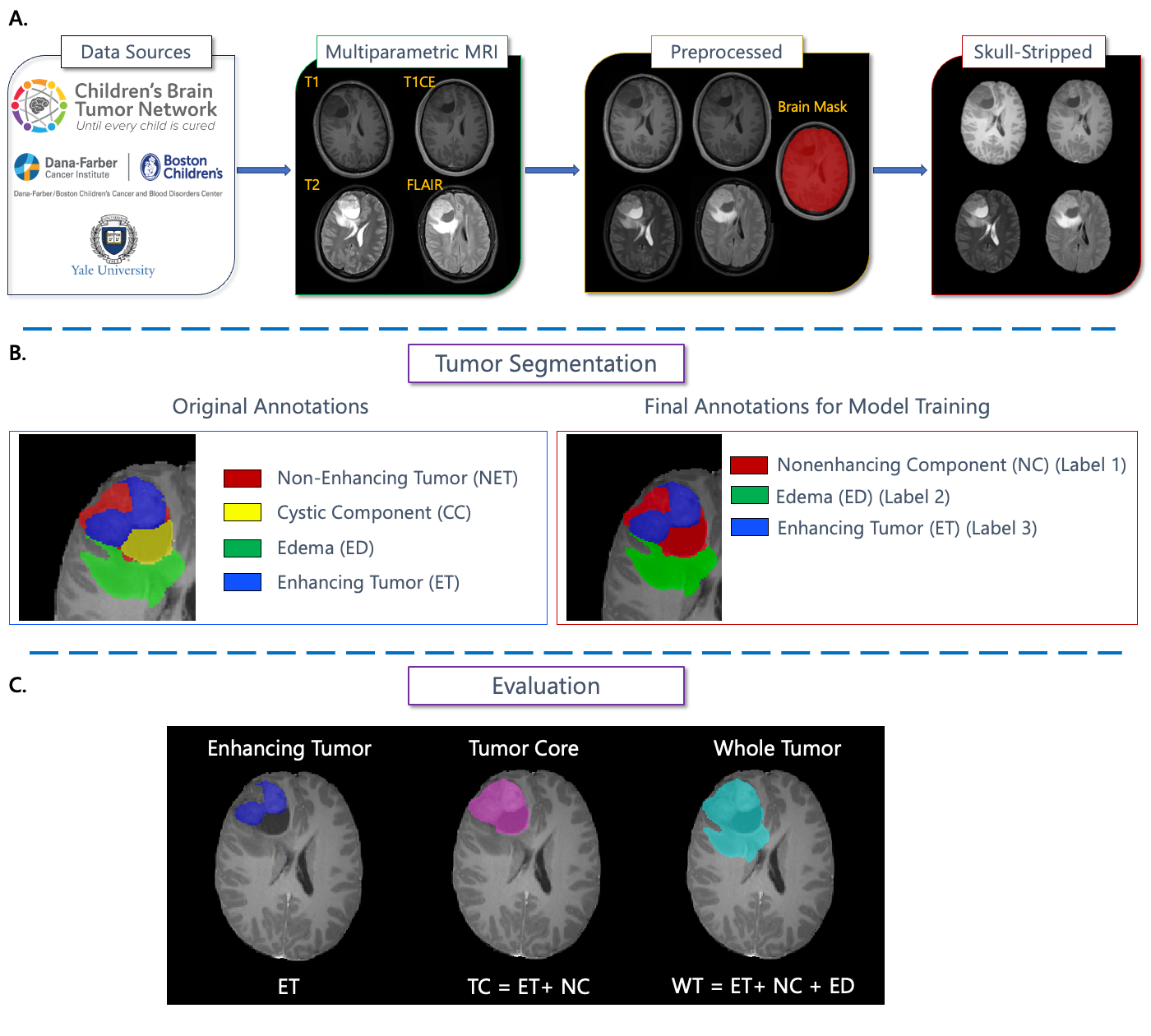
- Curated multi-institution pediatric MRI dataset with four sequences (T1, T1CE, T2, FLAIR) and ground-truth labels for ET, NC, CC, and ED (mapped to three labels for challenge).
- Pre-processing to 1 mm3 resolution, co-registration to template, and de-identification using pediatric skull-stripping.
- Participants trained on provided data; evaluation used lesionwise Dice and HD95 across ET, TC, WT with 24 test subjects.
- Ground truth segmentations refined by neuroradiologists and ASNR reviewers following standardized annotation guidelines.
Experimental results
Research questions
- RQ1How well can AI segmentation methods reproduce pediatric brain tumor subregions (ET, NC/TC, WT) on multi-institution mpMRI data?
- RQ2Do ensembles of state-of-the-art architectures (e.g., nnU-Net, Swin UNETR) outperform single-model approaches in pediatric tumor segmentation?
- RQ3What is the impact of pediatric-specific ground-truth annotation and preprocessing on cross-institution segmentation performance?
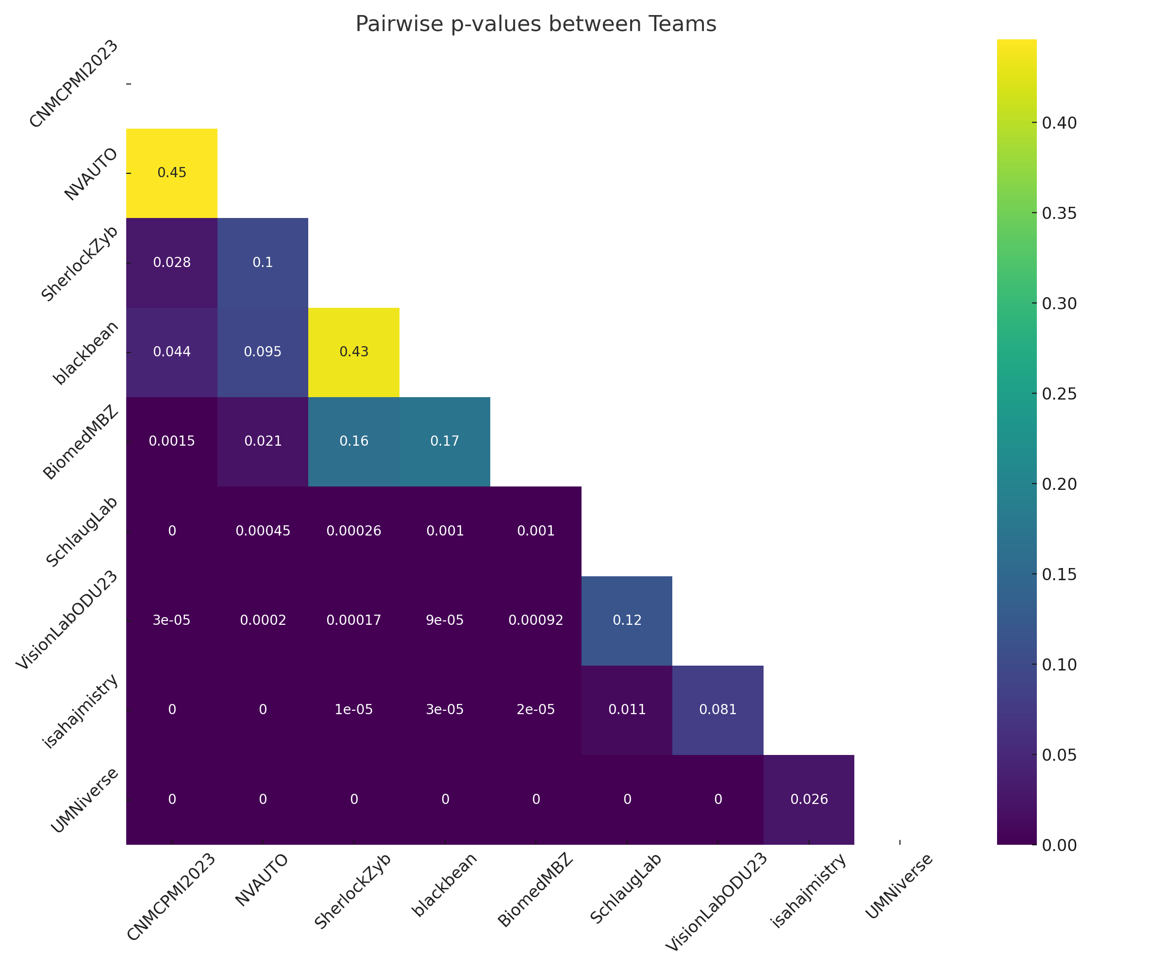
- RQ4Is there statistically significant performance difference between top teams on the BraTS-PEDs 2023 test set?
- RQ5Can ensembling and self-supervised pretraining improve robustness across heterogeneous pediatric MRI data?
Key findings
- Top approaches were ensembles of nnU-Net and Swin UNETR, Auto3DSeg, or nnU-Net with self-supervised pretraining.
- Mean Dice for WT around 0.83–0.84 and TC around 0.77–0.81 across top teams.
- ET segmentation showed more variability with Dice ~0.55–0.65, reflecting challenges with small or absent ET regions in DMG/DIPG.
- Ensembling may improve overall performance by combining strengths and mitigating weaknesses.
- Statistical significance of top-team differences was limited by small test cohort (n=24).
- WT segmentation was the most reliable across teams; ET remained the most challenging subregion.
Better researchstarts right now
From reading papers to final review, dramatically reduce your research time.
No credit card · Free plan available
This review was created by AI and reviewed by human editors.