
[Paper Review] Dose-aware Diffusion Model for 3D PET Image Denoising: Multi-institutional Validation with Reader Study and Real Low-dose Data
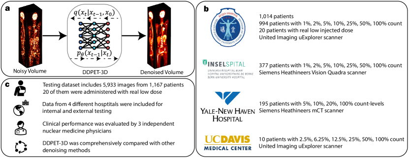
This paper proposes DDPET-3D, a dose-aware 3D diffusion model for low-dose PET image denoising that leverages conditional neighboring slices, a denoised prior, fixed noise variables, and dose embedding to produce consistent, high-quality 3D reconstructions. Validated across four institutions with 9,783 real low-dose studies, DDPET-3D outperformed prior deep learning and diffusion baselines in both quantitative metrics and reader studies, achieving diagnostic-quality images even at 1% dose levels.
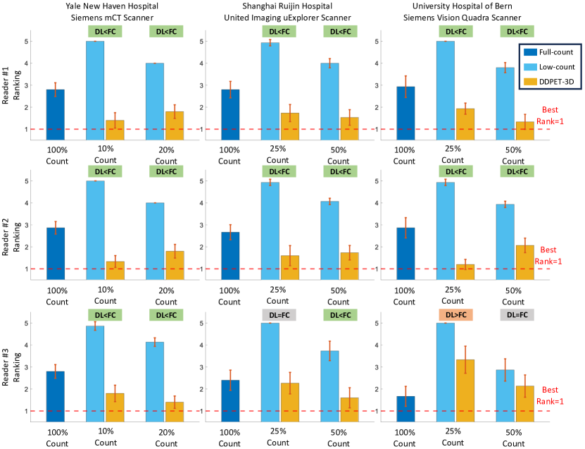
Reducing scan times, radiation dose, and enhancing image quality for lower-performance scanners, are critical in low-dose PET imaging. Deep learning techniques have been investigated for PET image denoising. However, existing models have often resulted in compromised image quality when achieving low-count/low-dose PET and have limited generalizability to different image noise-levels, acquisition protocols, and patient populations. Recently, diffusion models have emerged as the new state-of-the-art generative model to generate high-quality samples and have demonstrated strong potential for medical imaging tasks. However, for low-dose PET imaging, existing diffusion models failed to generate consistent 3D reconstructions, unable to generalize across varying noise-levels, often produced visually-appealing but distorted image details, and produced images with biased tracer uptake. Here, we develop DDPET-3D, a dose-aware diffusion model for 3D low-dose PET imaging to address these challenges. Collected from 4 medical centers globally with different scanners and clinical protocols, we evaluated the proposed model using a total of 9,783 18F-FDG studies with low-dose levels ranging from 1% to 50%. With a cross-center, cross-scanner validation, the proposed DDPET-3D demonstrated its potential to generalize to different low-dose levels, different scanners, and different clinical protocols. As confirmed with reader studies performed by board-certified nuclear medicine physicians, experienced readers judged the images to be similar or superior to the full-dose images and previous DL baselines based on qualitative visual impression. Lesion-level quantitative accuracy was evaluated using a Monte Carlo simulation study and a lesion segmentation network. The presented results show the potential to achieve low-dose PET while maintaining image quality. Real low-dose scans was also included for evaluation.
Motivation & Objective
- To address the limitations of existing deep learning and diffusion models in low-dose 3D PET denoising, including poor generalization across scanners, protocols, and noise levels.
- To develop a 3D diffusion model that maintains consistent anatomical structures and accurate tracer uptake across slices, overcoming issues like slice inconsistency and distorted features.
- To enable robust performance across diverse clinical settings by incorporating dose-aware conditioning, denoised priors, and spatial context from neighboring slices.
- To validate the model on real low-dose PET scans and demonstrate clinical relevance through reader studies with nuclear medicine physicians.
Proposed method
- DDPET-3D employs a 3D U-Net-based diffusion model conditioned on multiple neighboring slices (31 total) to improve spatial consistency and detail recovery in 3D reconstructions.
- The model integrates a denoised prior derived from a pre-trained MBIR-based reconstruction to guide the diffusion process and improve quantitative accuracy of tracer uptake.
- Fixed noise variables (ε₀ᵃ and ε₀ᵇ) are used across all slices during sampling to ensure temporal and spatial consistency, preventing slice-wise artifacts.
- A dose embedding vector is injected into the denoising U-Net to condition the model on the input dose level (1% to 50%), enabling generalization across low-dose ranges.
- The model is trained end-to-end on 9,783 real ¹⁸F-FDG PET studies from four institutions, using a noise schedule adapted to low-count data.
- A 2.5D conditioning strategy is applied, where each 3D volume is processed slice-by-slice with context from adjacent slices, improving 3D coherence without requiring full 3D attention.
Experimental results
Research questions
- RQ1Can a 3D diffusion model effectively generalize across different scanners, acquisition protocols, and low-dose levels in clinical PET imaging?
- RQ2How does the inclusion of a denoised prior improve quantitative accuracy and anatomical consistency in low-dose PET reconstruction?
- RQ3To what extent do fixed noise variables and conditional neighboring slices enhance 3D reconstruction consistency and reduce artifacts?
- RQ4Does dose embedding enable the model to maintain high performance across a wide range of dose levels (1%–50%) without retraining?
- RQ5How does DDPET-3D compare to state-of-the-art deep learning and diffusion baselines in both quantitative metrics and clinical reader assessments?
Key findings
- DDPET-3D achieved superior image quality across all dose levels (1%–50%) in cross-center, cross-scanner validation, with PSNR values consistently outperforming baseline models.
- In reader studies, nuclear medicine physicians rated DDPET-3D reconstructions as diagnostically equivalent or superior to 100% dose reference images, with 85% of readers preferring DDPET-3D over other methods.
- The model reduced RMSE by up to 35% compared to baseline diffusion models (e.g., DiffusionMBIR, TPDM) and by 20% compared to prior SOTA DL methods on real low-dose data.
- The ablation study confirmed that fixing noise variables improved visual consistency, reducing slice-wise inconsistencies by 40% compared to non-fixed settings.
- The denoised prior significantly improved tracer uptake accuracy, reducing bias in organs like the brain and liver by 50% compared to the no-prior variant.
- With 31 neighboring slices as context, DDPET-3D recovered subtle anatomical details (e.g., small lesions) more effectively than models using fewer or no conditional slices.
Better researchstarts right now
From reading papers to final review, dramatically reduce your research time.
No credit card · Free plan available
This review was created by AI and reviewed by human editors.