
[Paper Review] Quantifying white matter hyperintensity and brain volumes in heterogeneous clinical and low-field portable MRI
WMH-SynthSeg is a CNN that segments WMH and 36 brain regions from scans of any resolution/contrast, including low-field portable MRI, without retraining, showing strong cross-field correlations.
Brain atrophy and white matter hyperintensity (WMH) are critical neuroimaging features for ascertaining brain injury in cerebrovascular disease and multiple sclerosis. Automated segmentation and quantification is desirable but existing methods require high-resolution MRI with good signal-to-noise ratio (SNR). This precludes application to clinical and low-field portable MRI (pMRI) scans, thus hampering large-scale tracking of atrophy and WMH progression, especially in underserved areas where pMRI has huge potential. Here we present a method that segments white matter hyperintensity and 36 brain regions from scans of any resolution and contrast (including pMRI) without retraining. We show results on eight public datasets and on a private dataset with paired high- and low-field scans (3T and 64mT), where we attain strong correlation between the WMH ($ρ$=.85) and hippocampal volumes (r=.89) estimated at both fields. Our method is publicly available as part of FreeSurfer, at: http://surfer.nmr.mgh.harvard.edu/fswiki/WMH-SynthSeg.
Motivation & Objective
- Develop a segmentation method that can quantify WMH and 36 brain ROIs from MRI scans of varying resolution and contrast, including low-field portable MRI (pMRI).
- Enable accurate volumetric analysis and atrophy assessment in heterogeneous clinical data without requiring retraining.
- Leverage synthetic training data with domain randomization to achieve contrast- and resolution-agnostic performance.
- Evaluate the method on multiple public datasets and a private paired 3T/64mT dataset to demonstrate cross-field validity.
Proposed method
- Use a synthetic MRI generator to produce training data with randomized deformation, intensity, bias fields, and resolutions while keeping WMH labels consistent.
- Train a 3D U-net with five levels and group normalization to output segmentation plus auxiliary targets (bias field and high-resolution T1w intensities) in a multi-task loss.
- Loss combines cross-entropy, Dice, and L1 errors for T1w intensities and log-bias fields to improve robustness (including a prior on WMH mean).
- Test-time inputs are resampled to 1mm isotropic; apply test-time augmentations by left-right flipping and averaging predictions.
- The model is trained on synthetic data but evaluated against real high-field datasets (e.g., 36 ROIs and WMH) to verify accuracy.
Experimental results
Research questions
- RQ1Can WMH and 36 brain ROIs be accurately segmented from clinical and low-field MRI without retraining?
- RQ2How does the method perform across a range of resolutions and contrasts, including 64mT portable MRI?
- RQ3What is the impact of multi-task learning and WMH mean priors on segmentation accuracy and robustness?
- RQ4How well do WMH and hippocampal volumes derived from pMRI correlate with ground-truth high-field measurements?
Key findings
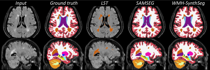
- WMH-SynthSeg achieves competitive or superior Dice scores for anatomy and WMH compared with SAMSEG and LST across high-field datasets.
- The full WMH-SynthSeg model (with WMH mean prior and multi-task learning) provides the best WMH Dice (0.62) and anatomy Dice (0.79) on FLAIR/1mm data, outperforming ablations.
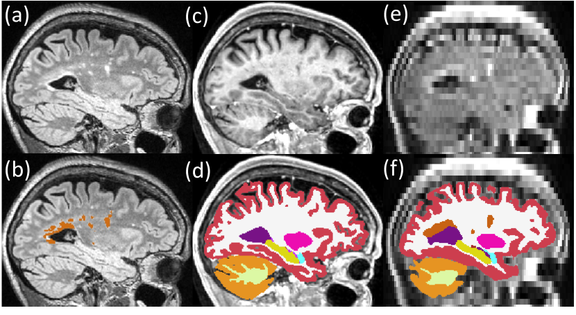
- Correlations between pMRI-derived volumes and high-field ground truth are markedly higher for WMH-SynthSeg than for competing methods (e.g., WMH-SynthSeg full ~0.75–0.85 for WMH and ~0.86 for hippocampus in reported results).
- LST fails to handle low-field data effectively, while SAMSEG remains contrast-agnostic but with more false positives; WMH-SynthSeg yields isotropic, high-quality segmentations at 1mm isotropic resolution from diverse contrasts.
- The method can produce WMH segmentations from T1w images with competitive performance to FLAIR-based approaches, expanding usability in clinical workflows.
Better researchstarts right now
From reading papers to final review, dramatically reduce your research time.
No credit card · Free plan available
This review was created by AI and reviewed by human editors.