
[Paper Review] The Rio Hortega University Hospital Glioblastoma dataset: a comprehensive collection of preoperative, early postoperative and recurrence MRI scans (RHUH-GBM)
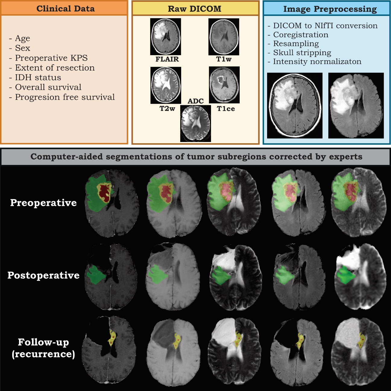
The RHUH-GBM dataset provides a comprehensive collection of multiparametric preoperative, early postoperative (within 72 hours), and recurrence MRI scans from 40 glioblastoma patients who underwent gross total or near-total resection. It includes expert-corrected tumor subregion segmentations, clinical data, molecular profiles, and survival outcomes, enabling advanced research in postoperative MRI analysis, recurrence modeling, and AI-driven segmentation and registration algorithms.
Glioblastoma, a highly aggressive primary brain tumor, is associated with poor patient outcomes. Although magnetic resonance imaging (MRI) plays a critical role in diagnosing, characterizing, and forecasting glioblastoma progression, public MRI repositories present significant drawbacks, including insufficient postoperative and follow-up studies as well as expert tumor segmentations. To address these issues, we present the "Río Hortega University Hospital Glioblastoma Dataset (RHUH-GBM)," a collection of multiparametric MRI images, volumetric assessments, molecular data, and survival details for glioblastoma patients who underwent total or near-total enhancing tumor resection. The dataset features expert-corrected segmentations of tumor subregions, offering valuable ground truth data for developing algorithms for postoperative and follow-up MRI scans.
Motivation & Objective
- To address the lack of public MRI datasets with early postoperative and recurrence scans for glioblastoma patients.
- To provide high-quality, expert-verified tumor subregion segmentations for preoperative, early postoperative, and recurrence time points.
- To support the development of AI-based registration and segmentation algorithms tailored for postoperative and follow-up MRI scans.
- To enable research into recurrence patterns and prognostic modeling in patients with gross total or near-total resection.
- To enhance the availability of multimodal MRI, clinical, and molecular data for glioblastoma research.
Proposed method
- Collected multiparametric MRI scans (T1w, T1ce, T2w, FLAIR, ADC) at three time points: preoperative, early postoperative (≤72 h), and recurrence.
- Selected 40 patients with WHO grade 4 astrocytoma who underwent gross total resection (GTR) or near-total resection (NTR) >95%.
- Performed image preprocessing using dcm2niix for DICOM-to-NIfTI conversion, FLIRT for linear registration to the SRI24 atlas, and Synthstrip for skull-stripping.
- Applied intensity Z-scoring normalization to standardize MRI signal intensities across subjects.
- Generated expert-corrected segmentations of enhancing tumor, non-enhancing tumor, and peritumoral T2/FLAIR hyperintensities at each time point.
- Integrated clinical, pathological, and treatment data including KPS, IDH status, Stupp protocol therapy, and survival outcomes.
Experimental results
Research questions
- RQ1How do tumor subregions evolve across preoperative, early postoperative, and recurrence MRI scans in glioblastoma patients with gross total resection?
- RQ2Can expert-annotated segmentations from early postoperative scans improve the accuracy of AI-based tumor segmentation and registration algorithms?
- RQ3What are the patterns of recurrence and peritumoral signal changes in patients with high extent of resection, as captured by multiparametric MRI?
- RQ4How do molecular markers (e.g., IDH status) and clinical factors correlate with recurrence timing and survival in this cohort?
- RQ5To what extent can the RHUH-GBM dataset improve the development of prognostic models for glioblastoma using multimodal MRI and clinical data?
Key findings
- The dataset comprises 40 glioblastoma patients with complete preoperative, early postoperative (within 72 hours), and recurrence MRI scans, all acquired at the same institution.
- All patients underwent gross total resection (GTR) or near-total resection (NTR) with >95% resection of the enhancing tumor volume.
- The median overall survival was 364 days, and the median progression-free survival was 198 days.
- The dataset includes expert-corrected segmentations of enhancing tumor, non-enhancing tumor, and peritumoral T2/FLAIR hyperintensities at all three time points.
- All patients received adjuvant chemotherapy and radiotherapy according to the Stupp protocol, with 72.5% receiving 60 Gy in 30 fractions via VMAT-IMRT-IGRT.
- The dataset is publicly available via The Cancer Imaging Archive (TCIA) and includes preprocessing code on GitHub for reproducibility.
Better researchstarts right now
From reading papers to final review, dramatically reduce your research time.
No credit card · Free plan available
This review was created by AI and reviewed by human editors.