
[论文解读] A Novel Deep Learning based Model for Erythrocytes Classification and Quantification in Sickle Cell Disease
本研究提出一种定制的18层深度卷积神经网络(CNN),用于使用来自镰状细胞病(SCD)患者的428张原始显微镜图像对三种红细胞形态——双凹盘状、椭圆形和镰状——进行分类和量化,生成包含10,377张单细胞图像的数据集。该模型实现了81%的准确率,优于基线模型,并通过SHAP和LIME提升可解释性,以支持临床决策。
The shape of erythrocytes or red blood cells is altered in several pathological conditions. Therefore, identifying and quantifying different erythrocyte shapes can help diagnose various diseases and assist in designing a treatment strategy. Machine Learning (ML) can be efficiently used to identify and quantify distorted erythrocyte morphologies. In this paper, we proposed a customized deep convolutional neural network (CNN) model to classify and quantify the distorted and normal morphology of erythrocytes from the images taken from the blood samples of patients suffering from Sickle cell disease ( SCD). We chose SCD as a model disease condition due to the presence of diverse erythrocyte morphologies in the blood samples of SCD patients. For the analysis, we used 428 raw microscopic images of SCD blood samples and generated the dataset consisting of 10, 377 single-cell images. We focused on three well-defined erythrocyte shapes, including discocytes, oval, and sickle. We used 18 layered deep CNN architecture to identify and quantify these shapes with 81% accuracy, outperforming other models. We also used SHAP and LIME for further interpretability. The proposed model can be helpful for the quick and accurate analysis of SCD blood samples by the clinicians and help them make the right decision for better management of SCD.
研究动机与目标
- 开发一种深度学习模型,用于自动分类和量化镰状细胞病(SCD)血液样本中的异常红细胞形态。
- 解决临床中手动评估红细胞形态带来的挑战,该方法耗时且易受观察者间差异影响。
- 创建一种可扩展、可解释的AI系统,支持临床医生在SCD早期诊断和治疗规划中的应用。
- 通过使模型能够适应其他具有形态学改变的血液系统疾病,拓展深度学习在SCD以外的应用潜力。
提出的方法
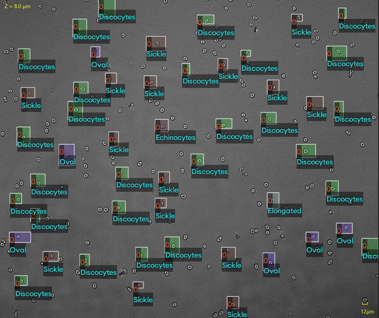
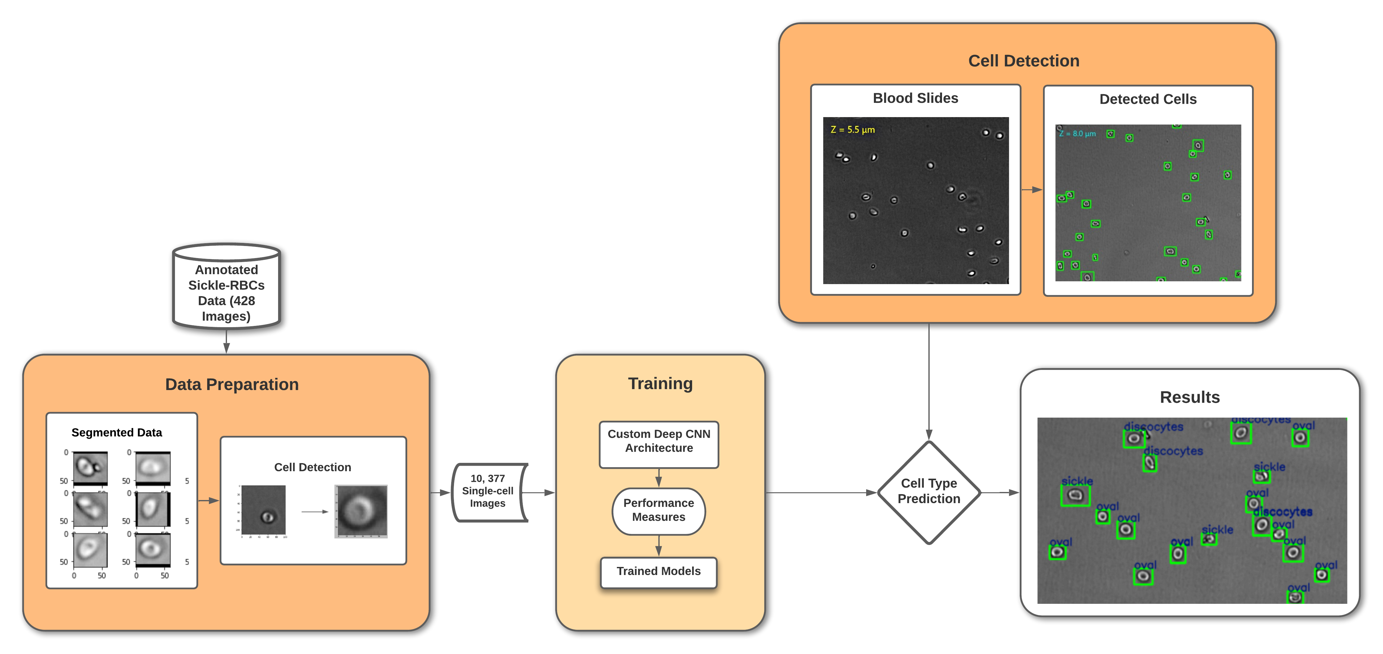
- 设计了一种定制的18层深度卷积神经网络(CNN),用于对红细胞形态进行多类别分类:双凹盘状、椭圆形和镰状。
- 该方法包括对原始显微镜图像进行预处理,并应用实例分割技术以分离出单个红细胞,从428张原始图像中生成10,377张单细胞图像。
- 评估了九种深度神经网络架构,包括迁移学习模型,使用微调后的LightGBM作为对比基准。
- 通过SHAP和LIME增强模型的可解释性,以解释预测结果,提高临床信任度和透明度。
- 应用数据增强和归一化技术以提升泛化能力并缓解类别不平衡问题。
- 使用70-15-15的数据集划分方式对模型进行训练和验证,评估指标包括准确率、精确率、召回率和F1分数。
实验结果
研究问题
- RQ1定制的深度CNN能否有效分类和量化SCD患者血液样本中三种不同的红细胞形态——双凹盘状、椭圆形和镰状?
- RQ2所提出的深度CNN模型在分类红细胞形态方面,与已建立的基线模型及LightGBM相比,性能如何?
- RQ3SHAP和LIME在多大程度上能够增强模型预测的可解释性,以提升临床可用性?
- RQ4该模型能否泛化到SCD以外的血液系统疾病中形态学异常的其他病理情况?
主要发现
- 所提出的18层深度CNN在分类红细胞形态方面实现了81%的准确率,优于所有基线模型和微调后的LightGBM。
- 该模型在所有三类(双凹盘状、椭圆形、镰状)中均表现出更优的精确率、召回率和F1分数,表明其具有稳健的多类别分类性能。
- SHAP和LIME分析显示,模型关注的特征包括细胞形状、曲率和膜不规则性,与临床专业知识一致。
- 包含10,377张单细胞图像的数据集为未来红细胞形态分类研究提供了宝贵且已标注的资源。
- 该模型在仅428张原始图像的有限数据集上表现良好,表明其在获取更大、更多样化的数据后具备强大的可扩展潜力。
- 本研究证明,深度学习可显著减少SCD中手动评估红细胞形态的时间和主观性,支持更快的临床决策。
更好的研究,从现在开始
从阅读论文到最终审阅,大幅缩短您的研究时间。
无需绑定信用卡
本解读由 AI 生成,并经人工编辑审核。