
[论文解读] AGMB-Transformer: Anatomy-Guided Multi-Branch Transformer Network for Automated Evaluation of Root Canal Therapy
本文提出 AGMB-Transformer,一种新颖的解剖结构引导的多分支 Transformer 网络,通过基于关键点的多项式曲线拟合进行分割以提取解剖特征,这些特征引导采用分层 Transformer 与组多头自注意力机制,从而提升自动化根管治疗评估中的分类准确率。该方法在 245 张图像的数据集上实现了 90.20% 的准确率,显著优于基线模型。
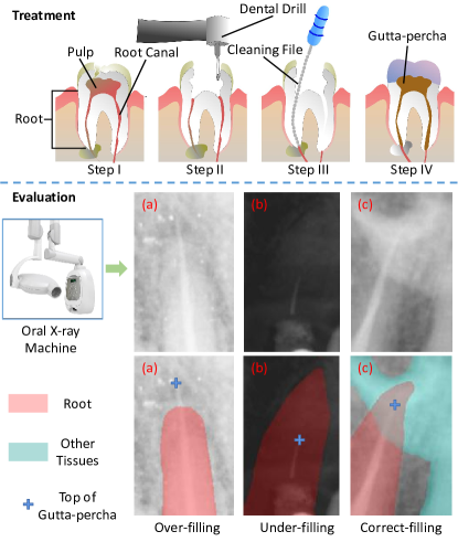
Accurate evaluation of the treatment result on X-ray images is a significant and challenging step in root canal therapy since the incorrect interpretation of the therapy results will hamper timely follow-up which is crucial to the patients' treatment outcome. Nowadays, the evaluation is performed in a manual manner, which is time-consuming, subjective, and error-prone. In this article, we aim to automate this process by leveraging the advances in computer vision and artificial intelligence, to provide an objective and accurate method for root canal therapy result assessment. A novel anatomy-guided multi-branch Transformer (AGMB-Transformer) network is proposed, which first extracts a set of anatomy features and then uses them to guide a multi-branch Transformer network for evaluation. Specifically, we design a polynomial curve fitting segmentation strategy with the help of landmark detection to extract the anatomy features. Moreover, a branch fusion module and a multi-branch structure including our progressive Transformer and Group Multi-Head Self-Attention (GMHSA) are designed to focus on both global and local features for an accurate diagnosis. To facilitate the research, we have collected a large-scale root canal therapy evaluation dataset with 245 root canal therapy X-ray images, and the experiment results show that our AGMB-Transformer can improve the diagnosis accuracy from 57.96% to 90.20% compared with the baseline network. The proposed AGMB-Transformer can achieve a highly accurate evaluation of root canal therapy. To our best knowledge, our work is the first to perform automatic root canal therapy evaluation and has important clinical value to reduce the workload of endodontists.
研究动机与目标
- 为解决牙髓治疗师进行手动、主观且耗时的根管治疗评估所面临的局限性。
- 开发一种利用医学影像与人工智能实现根管治疗结果自动、客观且准确分类的自动化方法。
- 克服 X 射线图像分析中的挑战,包括边界不清晰、图像质量差以及解剖结构遮挡等问题。
- 将解剖先验知识整合到深度学习中,以提升牙科影像中的诊断性能。
- 建立首个基于深度学习的根管治疗全流程自动化评估框架。
提出的方法
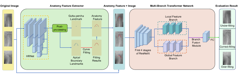
- 采用多项式曲线拟合分割策略,通过在根管 X 射线图像的关键点上拟合曲线来提取解剖特征。
- 将解剖特征与输入的 X 射线图像融合,并输入至带有专用分支融合模块的多分支 Transformer 架构中。
- 采用渐进式 Transformer 机制,对图像中的长距离全局依赖关系进行建模,增强上下文理解能力。
- 引入组多头自注意力机制(GMHSA),以高效捕捉局部空间模式并提升特征表示能力。
- 网络通过交叉熵损失进行端到端训练,实现对治疗结果(填充不足、填充过度或填充恰当)的多分类。
- 通过消融研究优化超参数,如特征块数量(θ)和多项式次数(δ)。
实验结果
研究问题
- RQ1基于解剖结构引导的特征是否能提升 X 射线图像中自动化根管治疗评估的准确率?
- RQ2与标准模型相比,具有渐进注意力机制和 GMHSA 的多分支 Transformer 架构在分类性能上表现如何?
- RQ3分支融合模块与多项式拟合次数的最佳配置是什么,以实现最大诊断准确率?
- RQ4所提出的方法是否可泛化至根管治疗以外的其他医学影像任务?
- RQ5在诊断中引入解剖先验在多大程度上可减少对高质量图像质量的依赖,并降低观察者间差异?
主要发现
- AGMB-Transformer 实现了 90.20% 的分类准确率,显著高于基线模型的 57.96%。
- 消融研究显示,最优配置为 θ = 64(64 个特征块)和 δ = 2(二次多项式拟合)。
- 在根管治疗数据集上,模型 AUC 达 95.63%,敏感度为 91.39%,特异度为 95.09%,F1 分数为 90.48%。
- 在 Plant Pathology 2020 数据集上评估时,AGMB-Transformer 达到 96.15% 的准确率,优于 ResNet50、ResNeXt50、GCNet50 和 BoTNet50。
- 分支融合模块能有效结合多个分支的特征,增强全局与局部特征学习能力。
- 该方法是首个将解剖结构引导的多分支 Transformer 应用于自动化根管治疗评估的方法,展现出强大的临床应用潜力。
更好的研究,从现在开始
从阅读论文到最终审阅,大幅缩短您的研究时间。
无需绑定信用卡
本解读由 AI 生成,并经人工编辑审核。