
[论文解读] Analysis of the 2024 BraTS Meningioma Radiotherapy Planning Automated Segmentation Challenge
本文介绍了2024年BraTS-MEN-RT挑战赛,这是一项大规模、多机构合作的项目,旨在为放射治疗计划中T1增强脑部MRI的脑膜瘤靶区体积开发自动化分割模型。利用单一原始分辨率的T1c序列和专家标注的轮廓,该挑战赛能够创建临床相关的开源模型,以减少轮廓勾画时间并提高治疗计划的一致性。
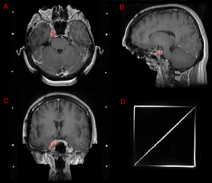
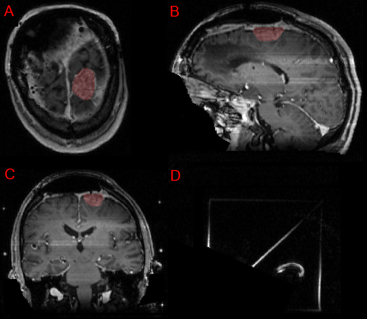
The 2024 Brain Tumor Segmentation Meningioma Radiotherapy (BraTS-MEN-RT) challenge aimed to advance automated segmentation algorithms using the largest known multi-institutional dataset of 750 radiotherapy planning brain MRIs with expert-annotated target labels for patients with intact or postoperative meningioma that underwent either conventional external beam radiotherapy or stereotactic radiosurgery. Each case included a defaced 3D post-contrast T1-weighted radiotherapy planning MRI in its native acquisition space, accompanied by a single-label "target volume" representing the gross tumor volume (GTV) and any at-risk post-operative site. Target volume annotations adhered to established radiotherapy planning protocols, ensuring consistency across cases and institutions, and were approved by expert neuroradiologists and radiation oncologists. Six participating teams developed, containerized, and evaluated automated segmentation models using this comprehensive dataset. Team rankings were assessed using a modified lesion-wise Dice Similarity Coefficient (DSC) and 95% Hausdorff Distance (95HD). The best reported average lesion-wise DSC and 95HD was 0.815 and 26.92 mm, respectively. BraTS-MEN-RT is expected to significantly advance automated radiotherapy planning by enabling precise tumor segmentation and facilitating tailored treatment, ultimately improving patient outcomes. We describe the design and results from the BraTS-MEN-RT challenge.
研究动机与目标
- 建立迄今为止公开可用的、由专家标注的脑膜瘤放射治疗计划MRIs最大数据集,用于自动化分割。
- 加速开发可鲁棒、可泛化的深度学习模型,以实现其在放射治疗计划中的临床集成。
- 通过聚焦于单一原始分辨率的T1增强MRI序列,简化模型的部署。
- 通过公开发布训练好的模型和数据,推动开放科学,以支持未来的研究和临床应用。
- 为未来针对多种脑肿瘤类型和多模态影像(例如CT、PET)的挑战赛奠定基础。
提出的方法
- 该挑战赛使用单一原始分辨率的T1增强脑部MRI序列作为分割模型的输入。
- 参赛者需使用来自多个机构的专家标注的真实值,对脑膜瘤的宏观肿瘤体积(GTV)进行分割。
- 所有数据均在机构审查委员会(IRB)批准下收集,并获得知情同意书的豁免。
- 数据集托管于Synapse平台,并将在挑战赛期间(2024年5月下旬至10月)公开发布。
- 使用标准分割指标(如Dice相似度系数(DSC)和Hausdorff距离)对模型进行评估。
- 通过多机构数据收集,促进联邦学习并提升模型的泛化能力。
实验结果
研究问题
- RQ1自动化深度学习模型能否在临床放射治疗计划中,仅使用单一T1增强MRI序列,实现对脑膜瘤宏观肿瘤体积的高精度分割?
- RQ2模型性能和泛化能力在不同机构和成像协议之间如何变化?
- RQ3开源的、多机构的数据集在多大程度上能促进临床可部署分割工具的开发?
- RQ4将自动化分割集成到现有放射治疗计划系统中,面临哪些关键技术与工作流程挑战?
- RQ5未来挑战赛如何能够超越脑膜瘤,涵盖其他脑肿瘤类型以及多模态影像(例如CT、PET)?
主要发现
- BraTS-MEN-RT挑战赛提供了目前已知最大的专家标注脑膜瘤放射治疗计划MRIs数据集,涵盖58家机构的数据。
- 数据集包含原始分辨率的T1增强MRI序列,可直接集成到临床工作流程中,无需预处理。
- 该挑战赛通过在挑战结束后于Synapse平台公开所有模型和数据,支持开放科学。
- 该倡议旨在减少手动轮廓勾画时间,并提高放射治疗计划中的一致性与可重复性。
- 该挑战赛为未来针对多模态影像(例如CT、PET)以及胶质瘤、听神经瘤等多样化脑肿瘤类型的科研工作奠定了基础。
- 模型和数据的公开发布有望加速开发鲁棒、可泛化的自动化分割工具,以支持临床应用。
更好的研究,从现在开始
从阅读论文到最终审阅,大幅缩短您的研究时间。
无需绑定信用卡
本解读由 AI 生成,并经人工编辑审核。