
[论文解读] Automatic Pulmonary Lobe Segmentation Using Deep Learning
该论文提出了一种用于CT扫描中自动肺叶分割的深度学习框架,采用结合Dice损失和焦点损失的混合损失函数,以解决类别不平衡和难以分类的体素问题。通过使用凸包裁剪进行预处理,并在50例人工标注的LUNA16扫描数据上进行训练,该模型在LUNA16数据集上实现了91.48%的平均Dice系数,在独立的Tianchi数据集上达到了94.17%,展现出在不同数据集间强大的泛化能力。
Pulmonary lobe segmentation is an important task for pulmonary disease related Computer Aided Diagnosis systems (CADs). Classical methods for lobe segmentation rely on successful detection of fissures and other anatomical information such as the location of blood vessels and airways. With the success of deep learning in recent years, Deep Convolutional Neural Network (DCNN) has been widely applied to analyze medical images like Computed Tomography (CT) and Magnetic Resonance Imaging (MRI), which, however, requires a large number of ground truth annotations. In this work, we release our manually labeled 50 CT scans which are randomly chosen from the LUNA16 dataset and explore the use of deep learning on this task. We propose pre-processing CT image by cropping region that is covered by the convex hull of the lungs in order to mitigate the influence of noise from outside the lungs. Moreover, we design a hybrid loss function with dice loss to tackle extreme class imbalance issue and focal loss to force model to focus on voxels that are hard to be discriminated. To validate the robustness and performance of our proposed framework trained with a small number of training examples, we further tested our model on CT scans from an independent dataset. Experimental results show the robustness of the proposed approach, which consistently improves performance across different datasets by a maximum of $5.87\%$ as compared to a baseline model.
研究动机与目标
- 开发一种在标注数据有限条件下的鲁棒深度学习框架,用于CT扫描中的肺叶分割。
- 通过结合Dice损失和焦点损失,解决肺叶分割中的极端类别不平衡问题。
- 通过凸包裁剪预处理提升模型在多样化CT数据集上的泛化能力。
- 公开发布50例来自LUNA16的、经人工标注的CT扫描数据集,以支持监督式肺叶分割研究。
提出的方法
- 通过提取肺部的凸包区域对CT扫描进行裁剪,以减少胸腔外区域的噪声影响。
- 采用混合损失函数,结合Dice损失以处理类别不平衡问题,以及焦点损失以聚焦于难以分类的体素。
- 通过随机平移、Z轴翻转和XY平面旋转进行数据增强,以提升模型泛化能力。
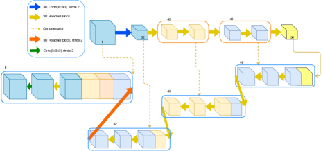
- 使用Adam优化器,以一个样本的批量大小,在300个周期内训练基于3D U-Net的深度卷积神经网络。
- 采用Dice系数评估性能,计算所有肺叶的逐叶Dice分数及平均Dice分数。
- 通过在独立的10例Tianchi数据集CT扫描上进行测试,验证模型的鲁棒性。
实验结果
研究问题
- RQ1仅使用50例人工标注的CT扫描,深度学习模型能否实现高性能的肺叶分割?
- RQ2在存在极端类别不平衡的情况下,结合Dice损失与焦点损失的混合损失函数在多大程度上提升了分割精度?
- RQ3通过凸包裁剪进行预处理,在多大程度上增强了模型在分布外CT扫描上的鲁棒性与性能?
- RQ4该模型在未参与训练的独立数据集(如Tianchi数据集)上的泛化能力如何?
- RQ5所提出的框架是否在肺叶分割任务中超越了现有最先进方法?
主要发现
- 所提出的框架在LUNA16测试集上实现了91.48%的最先进平均Dice系数,在Tianchi测试集上达到94.17%。
- 与仅使用Dice损失的基线模型相比,加入焦点损失后,LUNA16上的平均Dice系数提升了3.87%,Tianchi数据集上提升了3.95%。
- 与仅使用混合损失的模型相比,通过凸包裁剪预处理,LUNA16上的平均Dice系数进一步提升了0.54%,Tianchi数据集上提升了1.92%。
- 在两个数据集上,该模型相比先前最先进方法[7]的平均Dice系数最高提升了2.39%。
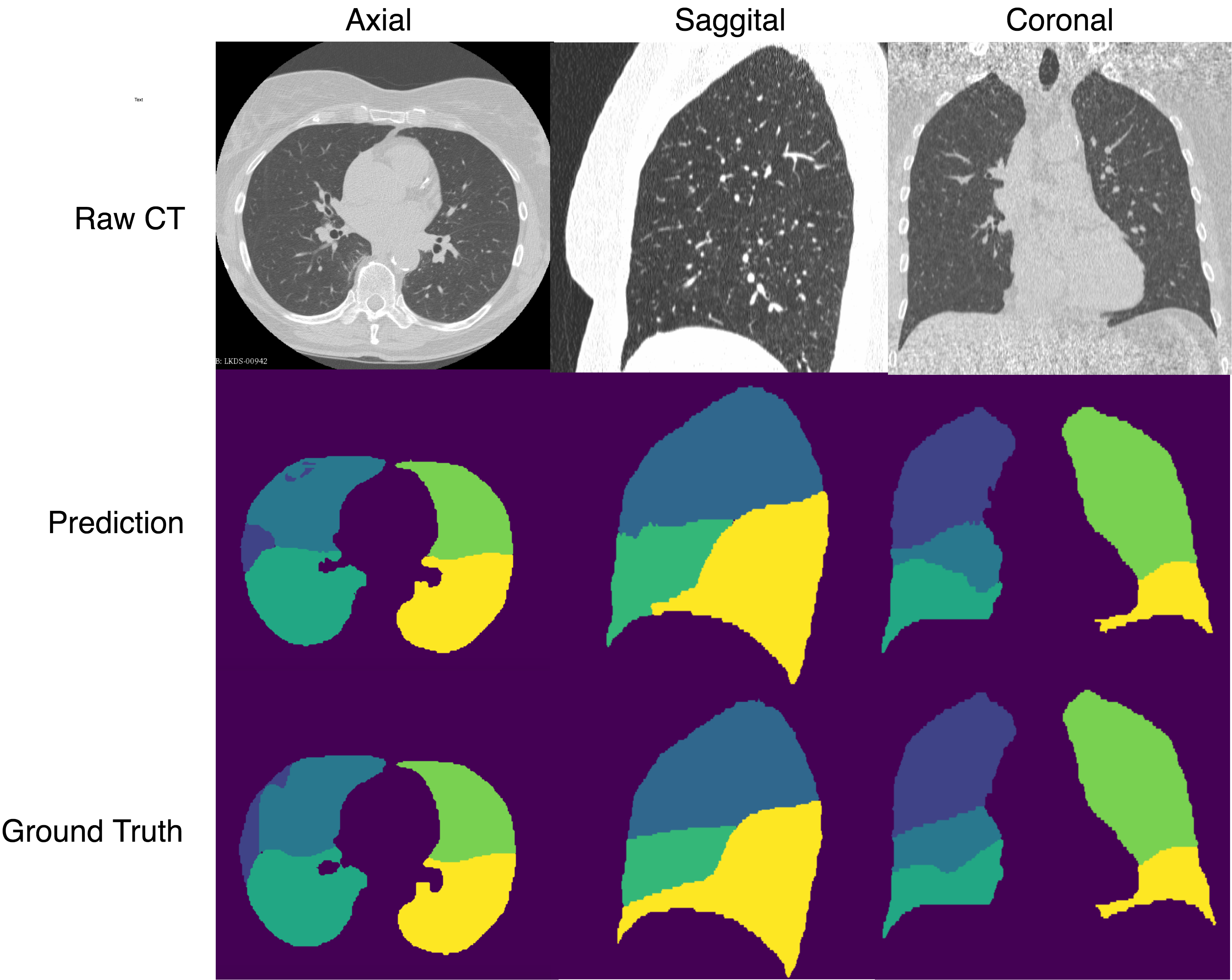
- 定性可视化结果表明,该模型能有效捕捉肺叶边界,并显著减少邻近裂隙区域的假阳性结果。
- 公开发布的50例来自LUNA16的人工标注CT扫描数据,为未来监督式肺叶分割研究提供了宝贵的基准。
更好的研究,从现在开始
从阅读论文到最终审阅,大幅缩短您的研究时间。
无需绑定信用卡
本解读由 AI 生成,并经人工编辑审核。