
[论文解读] Dose-aware Diffusion Model for 3D PET Image Denoising: Multi-institutional Validation with Reader Study and Real Low-dose Data
该论文提出 DDPET-3D,一种用于低剂量正电子发射断层扫描(PET)图像去噪的剂量感知3D扩散模型,通过利用条件相邻切片、去噪先验、固定噪声变量和剂量嵌入,生成一致且高质量的3D重建图像。在四个机构的9,783例真实低剂量研究中验证,DDPET-3D在定量指标和放射科医生评估中均优于先前的深度学习与扩散基线模型,即使在1%剂量水平下也能实现诊断级图像质量。
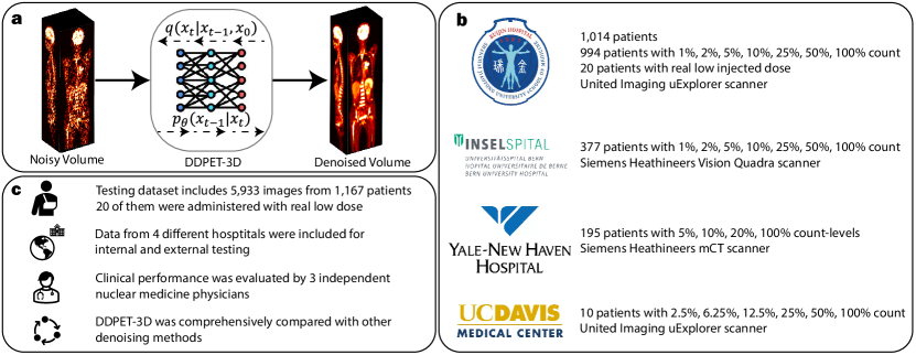
Reducing scan times, radiation dose, and enhancing image quality for lower-performance scanners, are critical in low-dose PET imaging. Deep learning techniques have been investigated for PET image denoising. However, existing models have often resulted in compromised image quality when achieving low-count/low-dose PET and have limited generalizability to different image noise-levels, acquisition protocols, and patient populations. Recently, diffusion models have emerged as the new state-of-the-art generative model to generate high-quality samples and have demonstrated strong potential for medical imaging tasks. However, for low-dose PET imaging, existing diffusion models failed to generate consistent 3D reconstructions, unable to generalize across varying noise-levels, often produced visually-appealing but distorted image details, and produced images with biased tracer uptake. Here, we develop DDPET-3D, a dose-aware diffusion model for 3D low-dose PET imaging to address these challenges. Collected from 4 medical centers globally with different scanners and clinical protocols, we evaluated the proposed model using a total of 9,783 18F-FDG studies with low-dose levels ranging from 1% to 50%. With a cross-center, cross-scanner validation, the proposed DDPET-3D demonstrated its potential to generalize to different low-dose levels, different scanners, and different clinical protocols. As confirmed with reader studies performed by board-certified nuclear medicine physicians, experienced readers judged the images to be similar or superior to the full-dose images and previous DL baselines based on qualitative visual impression. Lesion-level quantitative accuracy was evaluated using a Monte Carlo simulation study and a lesion segmentation network. The presented results show the potential to achieve low-dose PET while maintaining image quality. Real low-dose scans was also included for evaluation.
研究动机与目标
- 为解决现有深度学习与扩散模型在低剂量3D PET去噪中的局限性,包括在不同扫描仪、扫描协议和噪声水平下的泛化能力差的问题。
- 开发一种3D扩散模型,以保持切片间解剖结构的一致性并准确还原示踪剂摄取,克服切片不一致和特征失真等问题。
- 通过引入剂量感知条件、去噪先验和相邻切片的空间上下文,实现模型在多样化临床环境中的稳健性能。
- 在真实低剂量PET扫描上验证该模型,并通过核医学医生的读者研究展示其临床相关性。
提出的方法
- DDPET-3D 采用基于3D U-Net的扩散模型,通过条件化31个相邻切片来提升3D重建中的空间一致性和细节恢复能力。
- 模型整合了一个来自预训练MBIR重建方法的去噪先验,以引导扩散过程,提高示踪剂摄取的定量准确性。
- 在采样过程中,所有切片均使用固定的噪声变量(ε₀ᵃ 和 ε₀ᵇ),以确保时间与空间上的一致性,防止切片级伪影。
- 将剂量嵌入向量注入去噪U-Net中,以根据输入剂量水平(1%至50%)对模型进行条件化,实现对低剂量范围的泛化能力。
- 模型在来自四个机构的9,783例真实¹⁸F-FDG PET研究上进行端到端训练,采用适配于低计数数据的噪声调度。
- 采用2.5D条件化策略,即每个3D体积按切片逐个处理,并利用相邻切片的上下文信息,在无需完整3D注意力机制的前提下提升3D一致性。
实验结果
研究问题
- RQ13D扩散模型是否能有效泛化于不同扫描仪、采集协议和低剂量水平的临床PET成像?
- RQ2引入去噪先验在低剂量PET重建中如何提升定量准确性和解剖一致性?
- RQ3固定噪声变量与条件化相邻切片在多大程度上增强了3D重建的一致性并减少了伪影?
- RQ4剂量嵌入是否使模型能够在不重新训练的情况下,保持在1%–50%广泛剂量范围内的高性能?
- RQ5DDPET-3D在定量指标和临床读者评估中与最先进深度学习与扩散基线相比表现如何?
主要发现
- 在跨中心、跨扫描仪的验证中,DDPET-3D在所有剂量水平(1%–50%)下均实现了卓越的图像质量,PSNR值持续优于基线模型。
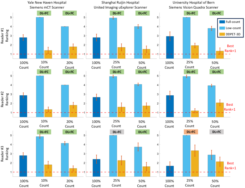
- 在读者研究中,核医学医生将DDPET-3D重建图像评为与100%剂量参考图像具有同等或更优的诊断价值,85%的读者更倾向于选择DDPET-3D而非其他方法。
- 与基线扩散模型(如DiffusionMBIR、TPDM)相比,DDPET-3D在真实低剂量数据上将RMSE降低了最多达35%;与先前SOTA深度学习方法相比,RMSE降低20%。
- 消融研究证实,固定噪声变量显著提升了视觉一致性,与非固定设置相比,切片间不一致性降低了40%。
- 去噪先验显著改善了示踪剂摄取的准确性,与无先验变体相比,脑部和肝脏等器官的偏差降低了50%。
- 在使用31个相邻切片作为上下文时,DDPET-3D比使用更少或无条件切片的模型更有效地恢复了细微解剖细节(如小病灶)。
更好的研究,从现在开始
从阅读论文到最终审阅,大幅缩短您的研究时间。
无需绑定信用卡
本解读由 AI 生成,并经人工编辑审核。