
[论文解读] MicroSSIM: Improved Structural Similarity for Comparing Microscopy Data
本文提出 MicroSSIM,一种专为显微镜图像评估而优化的结构相似性度量,解决了标准 SSIM 在低信噪比、高亮度、受偏移影响的显微镜数据中失效的问题。通过应用背景去除、图像下采样和自适应亮度缩放,MicroSSIM 降低了饱和效应,提升了对结构差异的敏感度,在真实显微镜数据集的去噪与分割任务中表现优于标准 SSIM。
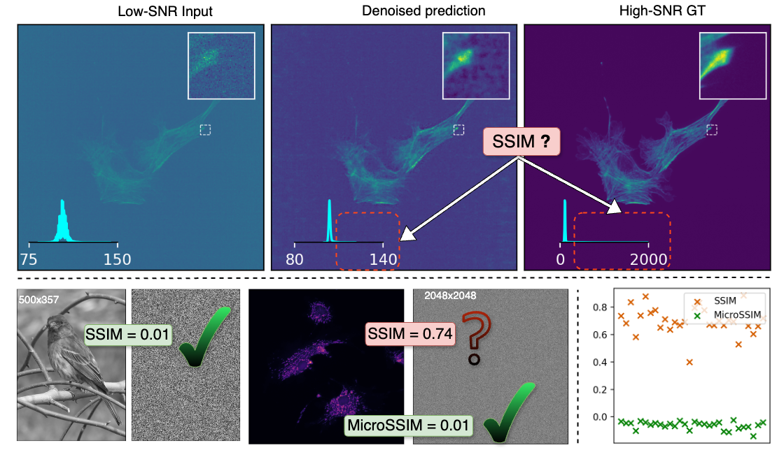
Microscopy is routinely used to image biological structures of interest. Due to imaging constraints, acquired images, also called as micrographs, are typically low-SNR and contain noise. Over the last few years, regression-based tasks like unsupervised denoising and splitting have found utility in working with such noisy micrographs. For evaluation, Structural Similarity (SSIM) is one of the most popular measures used in the field. For such tasks, the best evaluation would be when both low-SNR noisy images and corresponding high-SNR clean images are obtained directly from a microscope. However, due to the following three peculiar properties of the microscopy data, we observe that SSIM is not well suited to this data regime: (a) high-SNR micrographs have higher intensity pixels as compared to low-SNR micrographs, (b) high-SNR micrographs have higher intensity pixels than found in natural images, images for which SSIM was developed, and (c) a digitally configurable offset is added by the detector present inside the microscope which affects the SSIM value. We show that SSIM components behave unexpectedly when the prediction generated from low-SNR input is compared with the corresponding high-SNR data. We explain this by introducing the phenomenon of saturation, where SSIM components become less sensitive to (dis)similarity between the images. We propose an intuitive way to quantify this, which explains the observed SSIM behavior. We introduce MicroSSIM, a variant of SSIM, which overcomes the above-discussed issues. We justify the soundness and utility of MicroSSIM using theoretical and empirical arguments and show the utility of MicroSSIM on two tasks: unsupervised denoising and joint image splitting with unsupervised denoising. Since our formulation can be applied to a broad family of SSIM-based measures, we also introduce MicroMS3IM, a microscopy-specific variation of MS-SSIM.
研究动机与目标
- 为解决标准 SSIM 在评估显微镜图像时表现不佳的问题,原因在于显微镜数据具有高亮度、低信噪比和探测器偏移等独特特性。
- 识别并量化 SSIM 饱和现象,即在显微镜数据中相似度分数对结构差异变得不敏感。
- 开发一种新度量 MicroSSIM,通过针对性预处理和自适应缩放,校正亮度不匹配与偏移伪影。
- 将方法扩展至 MS-SSIM,提出 MicroMS3IM,用于显微镜应用中的多尺度评估。
- 在真实任务中验证该度量的优越性,如无监督去噪和联合图像分割。
提出的方法
- 提出 MicroSSIM 作为 SSIM 的改进形式,通过背景去除分离前景结构后再进行比较。
- 引入图像下采样以降低动态范围,减轻 SSIM 各分量中的饱和效应。
- 应用自适应亮度缩放因子 α,按数据集独立估计,以对齐低信噪比与高信噪比图像的亮度。
- 采用单一、基于数据集的 β(背景亮度)估计值,确保所有图像对之间亮度减法的一致性。
- 通过将相同的预处理与缩放流程应用于多尺度 SSIM,将方法扩展至 MS-SSIM,形成 MicroMS3IM。
- 采用现成的优化方法估计 α,确保实用性和与现有评估流程的兼容性。
实验结果
研究问题
- RQ1为何标准 SSIM 尽管被广泛使用,却在显微镜图像去噪性能评估中表现不可靠?
- RQ2显微镜数据中的高像素亮度与探测器引入的偏移如何扭曲 SSIM 分数?
- RQ3SSIM 饱和在多大程度上降低了对噪声与干净显微镜图像间结构差异的敏感度?
- RQ4预处理与缩放流程能否恢复 SSIM 在显微镜评估中的敏感性与可靠性?
- RQ5MicroSSIM 在真实去噪与分割基准测试中与 SSIM 和 MS-SSIM 相比表现如何?
主要发现
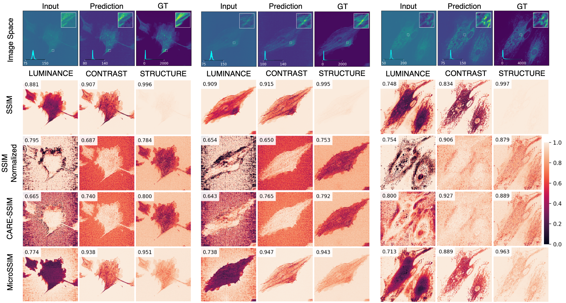
- MicroSSIM 显著降低了所有 SSIM 分量中的饱和因子 Δ,尤其在结构项中,提升了对图像差异的敏感度。
- 所提出的预处理(背景去除、下采样与 α 缩放)在对比高信噪比真实图像与纯噪声时,如预期产生更低的 SSIM 值,而标准 SSIM 则不然。
- MicroSSIM 对差异较大的图像对(如真实图像 vs. 均匀噪声)产生更低的 SSIM 值,同时对相似图像对(如真实图像 vs. 去噪预测)保持高分值,表现出正确的行为。
- 基于数据集的 β 估计确保了亮度减法的一致性与可靠性,防止 SSIM 分数被人为虚高。
- MicroMS3IM(多尺度扩展)相较于标准 MS-SSIM 表现更优,证实了该方法可推广至其他 SSIM 变体。
- 在 N2V 与 denoiSplit 模型上的实证结果表明,MicroSSIM 更能真实反映模型性能,对较差预测的分数下降更敏感、更具意义。
更好的研究,从现在开始
从阅读论文到最终审阅,大幅缩短您的研究时间。
无需绑定信用卡
本解读由 AI 生成,并经人工编辑审核。