[论文解读] The Brain Tumor Segmentation in Pediatrics (BraTS-PEDs) Challenge: Focus on Pediatrics (CBTN-CONNECT-DIPGR-ASNR-MICCAI BraTS-PEDs)
本论文描述 BraTS-PEDs 挑战,聚焦儿科脑肿瘤分割,详细说明数据、处理流程、注释工作流、评估与跨国际联盟的参与时间表。
Pediatric tumors of the central nervous system are the most common cause of cancer-related death in children. The five-year survival rate for high-grade gliomas in children is less than 20%. Due to their rarity, the diagnosis of these entities is often delayed, their treatment is mainly based on historic treatment concepts, and clinical trials require multi-institutional collaborations. Here we present the CBTN-CONNECT-DIPGR-ASNR-MICCAI BraTS-PEDs challenge, focused on pediatric brain tumors with data acquired across multiple international consortia dedicated to pediatric neuro-oncology and clinical trials. The CBTN-CONNECT-DIPGR-ASNR-MICCAI BraTS-PEDs challenge brings together clinicians and AI/imaging scientists to lead to faster development of automated segmentation techniques that could benefit clinical trials, and ultimately the care of children with brain tumors.
研究动机与目标
- 使用回顾性多机构 mpMRI 数据集对儿科脑肿瘤分割方法进行基准评估。
- 提供儿科肿瘤亚区的真实标签注释,以实现标准化评估。
- 促进临床医生与人工智能研究人员之间的协作,加速儿科神经肿瘤学自动分割工具的开发。
提出的方法
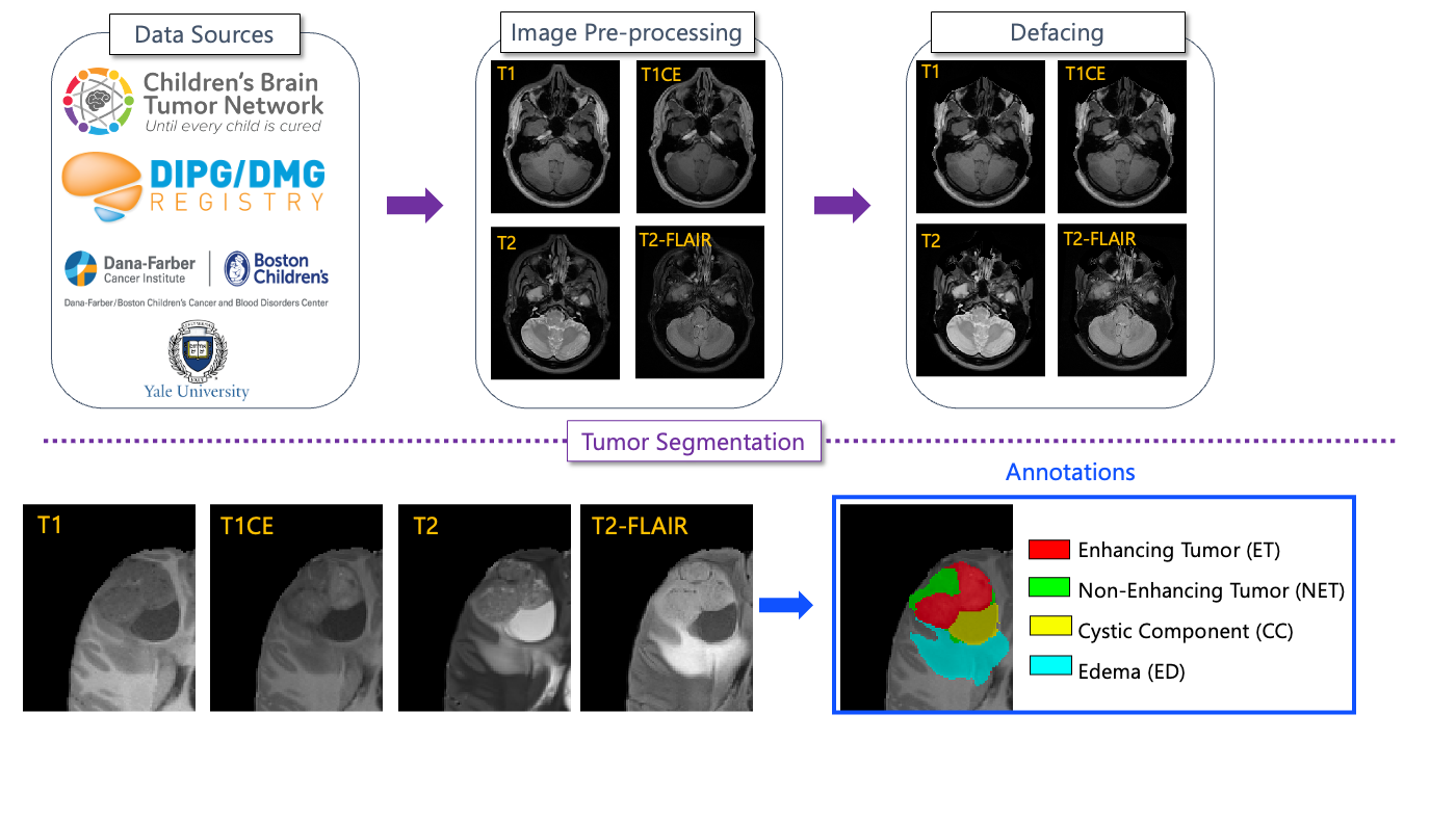
- 来自多个国际联盟(CBTN、CONNECT、DIPG Registry、ASNR)的数据聚合,形成464例儿科数据集。
- MRI 序列包括 T1、T1CE、T2 和 T2-FLAIR,且为未受治疗成像。
- 真实标签注释将肿瘤分成四个亚区:Enhancing Tumor (ET)、Nonenhancing Tumor (NET)、Cystic Component (CC) 和 Edema (ED)。
- 一款儿童自分割工具提供初步分割,经志愿神经放射学专家精炼并由主治神经放射科医生裁定。
- 预处理使用 BraTS Pipeline 与 CaPTk/FeTS 工具;为隐私对图像进行去识别化和匿名化。
- 评估聚焦于 ET、NC(ET/CC/坏死)和 Whole Tumor (WT);提交以容器化形式在由 MedPerf 提供支持的 Synapse 平台上进行评估。
实验结果
研究问题
- RQ1多机构的儿科 mpMRI 数据是否支持对儿科脑肿瘤的鲁棒自动分割?
- RQ2儿科肿瘤亚区(ET、NET、CC、ED)如何影响分割性能与治疗监测?
- RQ3标准化预处理和专家验证的真实标签对跨机构可重复性有何影响?
主要发现
- 回顾性多机构数据集包含464例儿科高等级胶质瘤,用于 BraTS-PEDs。
- 数据集包括四个 mpMRI 序列(T1、T1CE、T2、T2-FLAIR)和四个肿瘤亚区作为注释。
- 自动分割由神经放射学专家进行精炼,随后由具备资质的神经放射科医生审核后公开发布。
- 挑战采用容器化提交,在未见测试数据上通过 Synapse/MedPerf 平台进行评估。
- 本研究概述了在儿童脑肿瘤自动分割中常见的错误,并提供评估指南。
- 该挑战设计促进可重复性并与先前 BraTS 迭代进行比较,数据通过 CaPTk/FeTS 流程进行处理。
更好的研究,从现在开始
从论文设计到论文写作,大幅缩短您的研究时间。
无需绑定信用卡
本解读由 AI 生成,并经人工编辑审核。