
[Paper Review] Diffusion Modeling with Domain-conditioned Prior Guidance for Accelerated MRI and qMRI Reconstruction
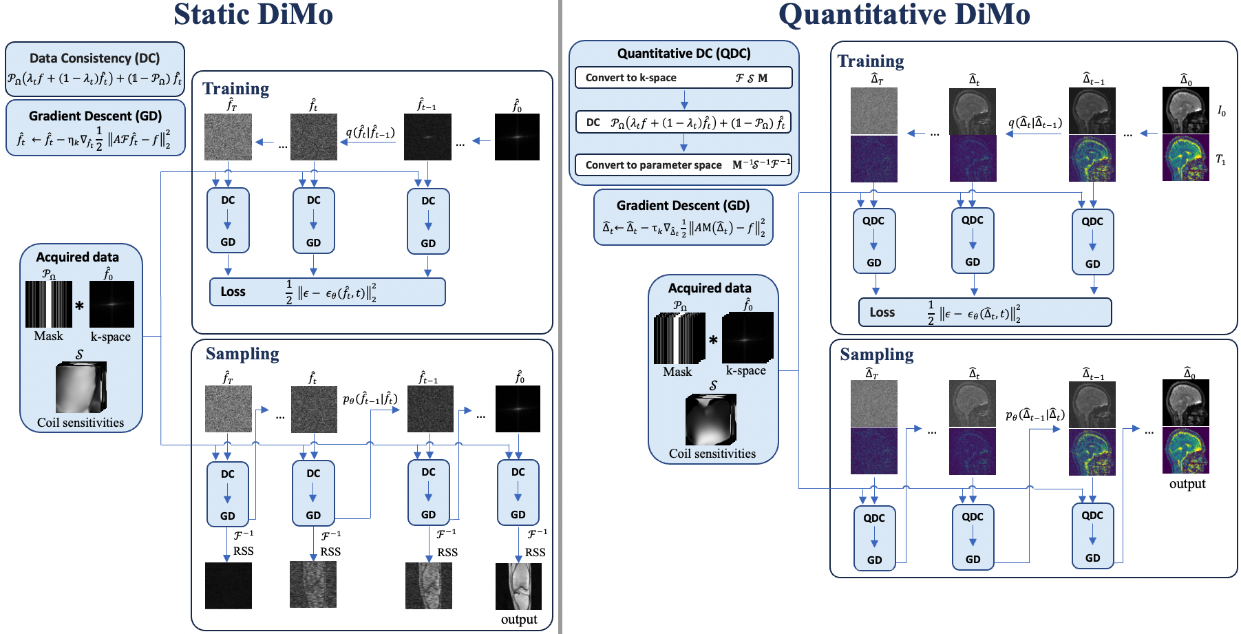
This paper proposes domain-conditioned diffusion models—Static DiMo for accelerated MRI and Quantitative DiMo for qMRI—by applying the diffusion process directly in the k-space and parameter domains, respectively, with physics-based priors and gradient-guided denoising. The method achieves state-of-the-art reconstruction accuracy and robustness at high acceleration factors, outperforming existing deep learning and optimization-based methods in both static and quantitative MRI.
This study introduces a novel approach for image reconstruction based on a diffusion model conditioned on the native data domain. Our method is applied to multi-coil MRI and quantitative MRI reconstruction, leveraging the domain-conditioned diffusion model within the frequency and parameter domains. The prior MRI physics are used as embeddings in the diffusion model, enforcing data consistency to guide the training and sampling process, characterizing MRI k-space encoding in MRI reconstruction, and leveraging MR signal modeling for qMRI reconstruction. Furthermore, a gradient descent optimization is incorporated into the diffusion steps, enhancing feature learning and improving denoising. The proposed method demonstrates a significant promise, particularly for reconstructing images at high acceleration factors. Notably, it maintains great reconstruction accuracy and efficiency for static and quantitative MRI reconstruction across diverse anatomical structures. Beyond its immediate applications, this method provides potential generalization capability, making it adaptable to inverse problems across various domains.
Motivation & Objective
- To address the limitations of existing diffusion models in MRI reconstruction, which often ignore physical constraints and operate in the image domain rather than the native data domain.
- To improve reconstruction accuracy and efficiency under high undersampling rates by embedding MRI physics (k-space encoding and signal modeling) directly into the diffusion process.
- To extend diffusion modeling to quantitative MRI (qMRI) for parameter mapping (e.g., T₁, I₀), a domain not previously explored with diffusion models.
- To enhance feature learning and denoising by integrating gradient descent optimization within the diffusion sampling steps.
- To develop a unified, generalizable framework applicable to diverse inverse problems in medical imaging beyond MRI.
Proposed method
- The forward and reverse diffusion processes are defined directly in the k-space domain for static MRI (Static DiMo), preserving the native frequency-domain structure of MRI data.
- For qMRI, the diffusion process is conditioned on the parameter domain (e.g., T₁, I₀), enabling reconstruction of quantitative maps from undersampled data.
- MRI physics—specifically k-space encoding and MR signal models—are embedded as data consistency components in the diffusion process to enforce physical plausibility.
- A gradient descent step is integrated into each diffusion sampling step, enhancing feature learning and improving denoising performance.
- The method uses a conditional diffusion model where the prior is guided by domain-specific embeddings derived from the underlying physics of MRI and qMRI.
- The framework is trained end-to-end using fully-sampled data, but during inference, it reconstructs high-quality images from highly undersampled k-space data.
Experimental results
Research questions
- RQ1Can diffusion modeling in the native data domain (k-space or parameter space) improve reconstruction accuracy and robustness in accelerated MRI compared to image-domain diffusion models?
- RQ2How does incorporating physics-based priors (k-space encoding and signal modeling) into the diffusion process affect reconstruction fidelity and generalization under distribution shifts?
- RQ3Can the integration of gradient descent within the diffusion sampling steps enhance feature learning and denoising without compromising image sharpness?
- RQ4Does the proposed method generalize to quantitative MRI reconstruction, where the goal is to recover tissue parameters like T₁ and I₀ from undersampled data?
- RQ5How does the method perform under extreme acceleration factors (e.g., 4×–6×), especially in terms of artifact suppression and edge preservation?
Key findings
- Quantitative DiMo achieved the lowest error in T₁ and I₀ mapping across all tested acceleration factors, with error maps showing the least deviation from fully-sampled references.
- At 4× 2D Poisson undersampling, Quantitative DiMo produced artifact-free T₁ and I₀ maps with superior structural detail, outperforming RELAX, LLR, and Model-TGV in sharpness and fidelity.
- The method maintained high reconstruction accuracy even at 6× acceleration with 1D Cartesian undersampling, where error and variance maps indicated increased uncertainty only at tissue boundaries.
- Ablation studies showed that increasing the number of sampling instances (from 10 to 100) reduced variance and improved mean image quality, confirming the method’s stability and uncertainty quantification capability.
- The integration of gradient descent within the diffusion steps significantly enhanced denoising while preserving fine anatomical details, as evidenced by reduced blurring and sharper edges in zoomed-in regions.
- The method demonstrated robustness across diverse anatomical structures, including frontal white matter, putamen, and thalamus, with consistent performance across multiple subjects and sequences.
Better researchstarts right now
From reading papers to final review, dramatically reduce your research time.
No credit card · Free plan available
This review was created by AI and reviewed by human editors.