
[논문 리뷰] BraTS-PEDs: Results of the Multi-Consortium International Pediatric Brain Tumor Segmentation Challenge 2023
이 논문은 BraTS-PEDs 2023을 제시하며, 소아 중심 BraTS 챌린지로Dataset, 평가 프로토콜, 참가 방법, 그리고 소아 고등형 교종에 대한 벤치마크된 분할 성능을 자세히 다룬다.
Pediatric central nervous system tumors are the leading cause of cancer-related deaths in children. The five-year survival rate for high-grade glioma in children is less than 20%. The development of new treatments is dependent upon multi-institutional collaborative clinical trials requiring reproducible and accurate centralized response assessment. We present the results of the BraTS-PEDs 2023 challenge, the first Brain Tumor Segmentation (BraTS) challenge focused on pediatric brain tumors. This challenge utilized data acquired from multiple international consortia dedicated to pediatric neuro-oncology and clinical trials. BraTS-PEDs 2023 aimed to evaluate volumetric segmentation algorithms for pediatric brain gliomas from magnetic resonance imaging using standardized quantitative performance evaluation metrics employed across the BraTS 2023 challenges. The top-performing AI approaches for pediatric tumor analysis included ensembles of nnU-Net and Swin UNETR, Auto3DSeg, or nnU-Net with a self-supervised framework. The BraTSPEDs 2023 challenge fostered collaboration between clinicians (neuro-oncologists, neuroradiologists) and AI/imaging scientists, promoting faster data sharing and the development of automated volumetric analysis techniques. These advancements could significantly benefit clinical trials and improve the care of children with brain tumors.
연구 동기 및 목표
- 다기관 mpMRI 데이터에서 소아 뇌종양의 자동 용적 분할을 벤치마크하고 표준화한다.
- 소아 종양 부분 영역의 실제 표준 라벨과 공정한 다중 센터 평가 프레임워크를 제공한다.
- 주로 U-Net 기반의 다양한 AI 접근법을 WT, TC, ET 분할에 대해 BraTS-PEDs 데이터에서 비교한다.
제안 방법
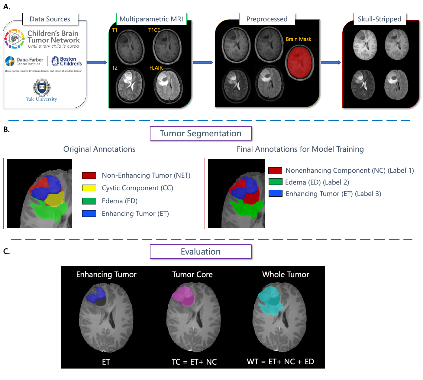
- 네 가지 시퀀스(T1, T1CE, T2, FLAIR)와 ET, NC, CC, ED의 실제 표준 라벨을 갖춘 다기관 소아 MRI 데이터셋을 큐레이션했다(챌린지용으로 3개의 라벨로 매핑).
- 해상도 1 mm3로 전처리하고 템플릿에 공동 정합(co-registration)하며 소아 두개골 제거를 통한 비식별화(De-identification) 절차를 적용했다.
- 참가자는 제공된 데이터로 학습했고, 평가에는 ET, TC, WT에 대해 24명의 테스트 피험자에서 병소별 Dice 및 HD95를 사용했다.
- 정상화된 주석 지침에 따라 뇌 신경영상 전문의와 ASNR 심사위원이 실제 라벨 분할을 정제했다.
실험 결과
연구 질문
- RQ1다기관 mpMRI 데이터에서 AI 분할 방법이 소아 뇌종양의 부분 영역(ET, NC/TC, WT)을 얼마나 잘 재현할 수 있는가?
- RQ2최신 아키텍처(nnU-Net, Swin UNETR 등)의 앙상블이 소아 종양 분할에서 단일 모델 접근법을 능가하는가?
- RQ3소아 특화 실제 표준 라벨링 및 전처리가 다기관 간 분할 성능에 어떤 영향을 주는가?
- RQ4BraTS-PEDs 2023 테스트 세트에서 최상위 팀 간 성능 차이가 통계적으로 유의한가?
- RQ5앙상블링과 자기지도 사전학습이 이질적인 소아 MRI 데이터에서 로버스트니스를 향상시킬 수 있는가?
주요 결과
- 최고의 접근 방식은 nnU-Net과 Swin UNETR의 앙상블, Auto3DSeg, 또는 자기지도 사전학습이 포함된 nnU-Net이었다.
- 상위 팀들 간 WT의 평균 Dice는 약 0.83–0.84, TC는 약 0.77–0.81 수준이었다.
- ET 분할은 Dice가 약 0.55–0.65로 변동성이 더 컸는데, 이는 DMG/DIPG에서 ET 영역이 작거나 부재한 경우를 반영한다.
- 앙상블은 강점을 결합하고 약점을 보완하여 전반적인 성능을 향상시킬 수 있다.
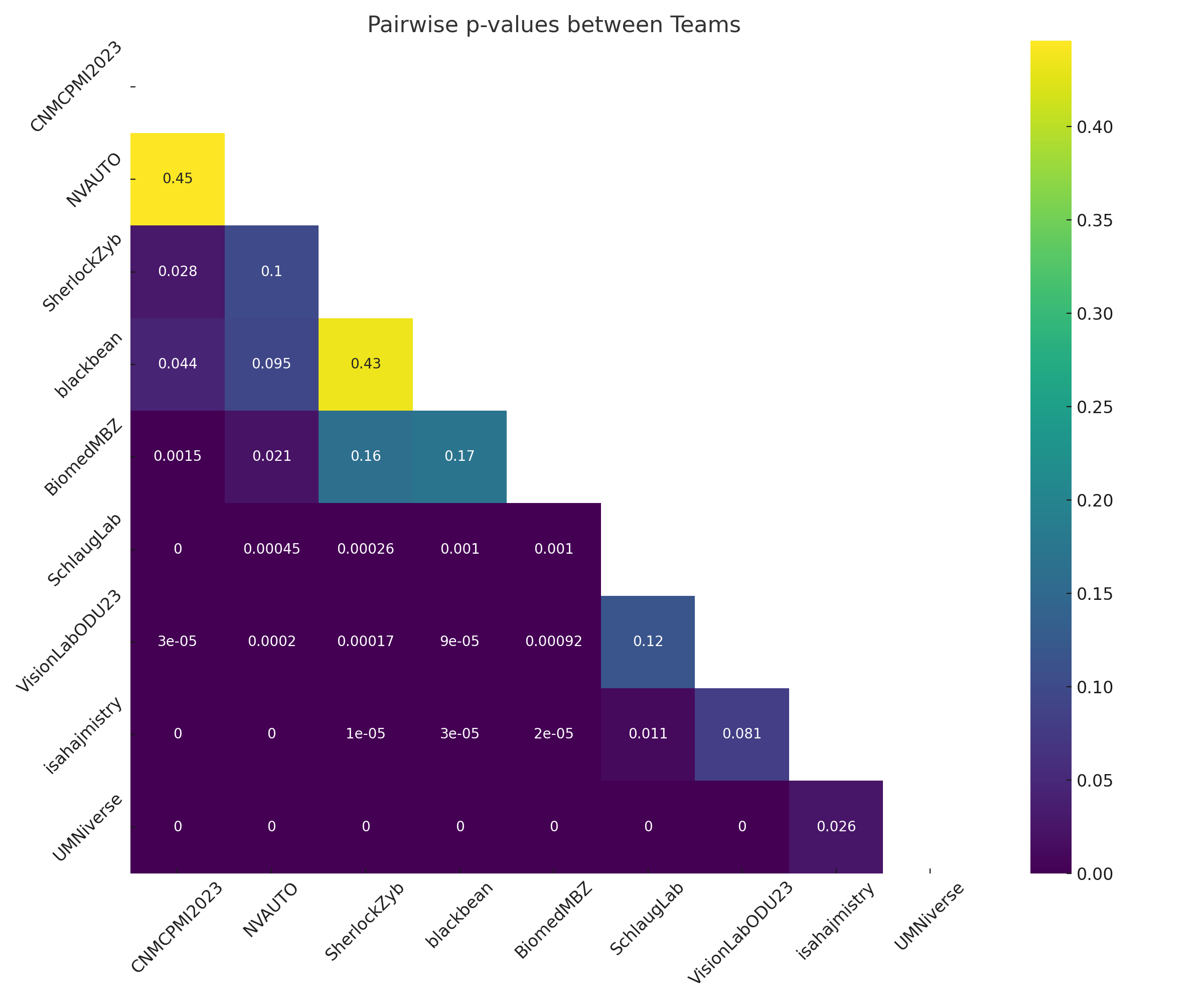
- 상위 팀 간의 통계적 유의성은 작은 테스트 코호트(n=24)로 인해 제한적이었다.
- WT 분할은 팀들 간에 가장 신뢰도가 높았지만 ET가 여전히 가장 도전적인 부분 영역이었다.
더 나은 연구,지금 바로 시작하세요
논문 읽기부터 검토까지, 연구 시간을 획기적으로 줄여보세요.
카드 등록 없음 · 무료 플랜 제공
이 리뷰는 AI가 만들고, 인간 에디터가 검토했습니다.