
[논문 리뷰] Dose-aware Diffusion Model for 3D PET Image Denoising: Multi-institutional Validation with Reader Study and Real Low-dose Data
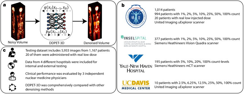
이 논문은 저선량 PET 영상의 노이즈 제거를 위한 복잡한 3D 확산 모델인 DDPET-3D를 제안한다. 이 모델은 조건부로 인접한 슬라이스, 노이즈 제거 전의 사전 정보, 고정된 노이즈 변수, 선량 임bedding를 활용하여 일관되고 고품질의 3D 재구성을 생성한다. 9,783건의 실제 저선량 연구 데이터를 활용해 4개 기관에서 검증된 결과, DDPET-3D는 정량적 지표와 독자 평가 모두에서 기존의 딥러닝 및 확산 기반 모델들을 능가했으며, 선량이 1% 수준일 경우에도 진단 수준의 영상을 확보했다.
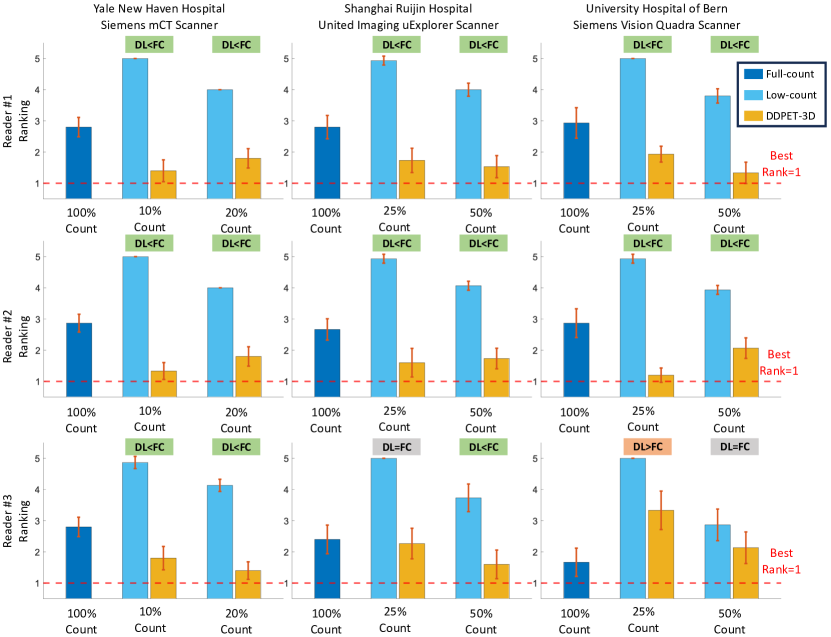
Reducing scan times, radiation dose, and enhancing image quality for lower-performance scanners, are critical in low-dose PET imaging. Deep learning techniques have been investigated for PET image denoising. However, existing models have often resulted in compromised image quality when achieving low-count/low-dose PET and have limited generalizability to different image noise-levels, acquisition protocols, and patient populations. Recently, diffusion models have emerged as the new state-of-the-art generative model to generate high-quality samples and have demonstrated strong potential for medical imaging tasks. However, for low-dose PET imaging, existing diffusion models failed to generate consistent 3D reconstructions, unable to generalize across varying noise-levels, often produced visually-appealing but distorted image details, and produced images with biased tracer uptake. Here, we develop DDPET-3D, a dose-aware diffusion model for 3D low-dose PET imaging to address these challenges. Collected from 4 medical centers globally with different scanners and clinical protocols, we evaluated the proposed model using a total of 9,783 18F-FDG studies with low-dose levels ranging from 1% to 50%. With a cross-center, cross-scanner validation, the proposed DDPET-3D demonstrated its potential to generalize to different low-dose levels, different scanners, and different clinical protocols. As confirmed with reader studies performed by board-certified nuclear medicine physicians, experienced readers judged the images to be similar or superior to the full-dose images and previous DL baselines based on qualitative visual impression. Lesion-level quantitative accuracy was evaluated using a Monte Carlo simulation study and a lesion segmentation network. The presented results show the potential to achieve low-dose PET while maintaining image quality. Real low-dose scans was also included for evaluation.
연구 동기 및 목표
- 기존의 딥러닝 및 확산 모델이 저선량 3D PET 노이즈 제거에서 겪는 한계, 즉 다양한 스크래너, 촬영 프로토콜, 노이즈 수준 간의 일반화 능력 부족 문제를 해결하기 위해.
- 슬라이스 간 일관성과 정확한 트레이서 집합을 유지하면서도, 슬라이스 불일치 및 왜곡된 특징 등의 문제를 해결하는 3D 확산 모델을 개발하기 위해.
- 선량 인지 조건화, 노이즈 제거 사전 정보, 인접 슬라이스의 공간적 맥락을 통합함으로써 다양한 임상 환경에서의 견고한 성능을 확보하기 위해.
- 실제 저선량 PET 스캔에서 모델을 검증하고, 핵의학의 전문의들이 참여하는 독자 평가를 통해 임상적 관련성을 입증하기 위해.
제안 방법
- DDPET-3D는 31개의 총 인접 슬라이스를 조건부로 사용하는 3D U-Net 기반의 확산 모델을 활용하여 3D 재구성에서 공간 일관성과 세부 사항 복구를 향상시킨다.
- 모델은 사전에 훈련된 MBIR 기반 재구성으로부터 유도된 노이즈 제거 사전 정보를 통합하여 확산 과정을 안내하고 트레이서 집합의 정량적 정확도를 향상시킨다.
- 모든 슬라이스에서 샘플링 중 고정된 노이즈 변수(ε₀ᵃ 및 ε₀ᵇ)를 사용하여 시간적·공간적 일관성을 확보하고 슬라이스 단위의 아티팩트를 방지한다.
- 선량 임bedding 벡터를 노이즈 제거 U-Net에 통합하여 입력 선량 수준(1%에서 50%)에 따라 조건화함으로써 저선량 범위 전반에서의 일반화를 가능하게 한다.
- 모델는 4개 기관의 9,783건의 실제 ¹⁸F-FDG PET 연구 데이터를 기반으로 엔드 투 엔드로 훈련되었으며, 저계수 데이터에 적합한 노이즈 스케줄을 사용했다.
- 2.5D 조건화 전략을 적용하여 각 3D 볼륨이 인접 슬라이스의 맥락을 활용해 슬라이스 단위로 처리되며, 전체 3D 어텐션을 요구하지 않으면서도 3D 일관성을 향상시켰다.
실험 결과
연구 질문
- RQ1다양한 스크래너, 촬영 프로토콜, 저선량 수준에서 임상 PET 영상에서 효과적으로 일반화할 수 있는 3D 확산 모델은 가능한가?
- RQ2노이즈 제거 사전 정보의 포함이 저선량 PET 재구성에서 정량적 정확도와 해부학적 일관성에 어떤 영향을 미치는가?
- RQ3고정된 노이즈 변수와 조건부 인접 슬라이스의 포함이 3D 재구성의 일관성 향상과 아티팩트 감소에 얼마나 기여하는가?
- RQ4선량 임bedding는 재학습 없이도 넓은 선량 수준 범위(1%–50%)에서 높은 성능을 유지하는 데 기여하는가?
- RQ5정량적 지표와 임상 독자 평가 모두에서 DDPET-3D는 최신의 딥러닝 및 확산 기반 모델들과 비교해 어떤가?
주요 결과
- 교차 기관, 교차 스크래너 검증에서 DDPET-3D는 모든 선량 수준(1%–50%)에서 뛰어난 영상 품질을 확보했으며, PSNR 값이 기준 모델들을 일관되게 뛰어넘었다.
- 독자 평가에서 핵의학의 전문의들은 DDPET-3D 재구성 영상을 100% 선량 기준 영상과 진단상 동등하거나 우수한 것으로 평가했으며, 85%의 독자들이 DDPET-3D를 다른 방법보다 선호했다.
- 기존의 기준 확산 모델들(DiffusionMBIR, TPDM)에 비해 실제 저선량 데이터에서 RMSE를 최대 35% 감소시켰고, 이전의 최고 수준의 딥러닝 방법에 비해도 20% 감소시켰다.
- 제거 분석 결과, 노이즈 변수를 고정함으로써 시각적 일관성이 향상되어, 비고정 설정 대비 슬라이스 간 불일치를 40% 감소시켰다.
- 노이즈 제거 사전 정보의 영향은 트레이서 집합 정확도를 크게 향상시켰으며, 뇌 및 간과 같은 장기에서의 편향을 비사전 정보 버전 대비 50% 감소시켰다.
- 31개의 인접 슬라이스를 맥락으로 사용함으로써 DDPET-3D는 더 작은 병변과 같은 미세한 해부학적 세부 사항을 더 효과적으로 복구했으며, 적은 수의 조건 슬라이스나 조건 없이 사용하는 모델들보다 뛰어난 성능을 보였다.
더 나은 연구,지금 바로 시작하세요
논문 읽기부터 검토까지, 연구 시간을 획기적으로 줄여보세요.
카드 등록 없음 · 무료 플랜 제공
이 리뷰는 AI가 만들고, 인간 에디터가 검토했습니다.