
[論文レビュー] Automatic Segmentation of Organs-at-Risk from Head-and-Neck CT using Separable Convolutional Neural Network with Hard-Region-Weighted Loss
本稿では、頭頸部CTスキャンにおける臓器リスク領域(OARs)の正確で不確実性を考慮したセグメンテーションを実現するため、分離可能畳み込みとハード領域重み付き損失を組み合わせた3D-SepNetフレームワークを提案する。セグメンテーション線形関数(SLF)を用いたマルチオルガニズム可視化と、不確実性推定のためのアンサンブル学習を統合することで、重み付き平均Diceスコア80.52%および95%ハウスドルフ距離3.043 mmを達成し、パラメータ数を1/3に抑えた3D UNetを上回る性能を発揮した。
Nasopharyngeal Carcinoma (NPC) is a leading form of Head-and-Neck (HAN) cancer in the Arctic, China, Southeast Asia, and the Middle East/North Africa. Accurate segmentation of Organs-at-Risk (OAR) from Computed Tomography (CT) images with uncertainty information is critical for effective planning of radiation therapy for NPC treatment. Despite the stateof-the-art performance achieved by Convolutional Neural Networks (CNNs) for automatic segmentation of OARs, existing methods do not provide uncertainty estimation of the segmentation results for treatment planning, and their accuracy is still limited by several factors, including the low contrast of soft tissues in CT, highly imbalanced sizes of OARs and large inter-slice spacing. To address these problems, we propose a novel framework for accurate OAR segmentation with reliable uncertainty estimation. First, we propose a Segmental Linear Function (SLF) to transform the intensity of CT images to make multiple organs more distinguishable than existing methods based on a simple window width/level that often gives a better visibility of one organ while hiding the others. Second, to deal with the large inter-slice spacing, we introduce a novel 2.5D network (named as 3D-SepNet) specially designed for dealing with clinic HAN CT scans with anisotropic spacing. Thirdly, existing hardness-aware loss function often deal with class-level hardness, but our proposed attention to hard voxels (ATH) uses a voxel-level hardness strategy, which is more suitable to dealing with some hard regions despite that its corresponding class may be easy. Our code is now available at https://github.com/HiLab-git/SepNet.
研究の動機と目的
- 放射線治療計画における頭頸部CTスキャンにおける低コントラストな軟部組織と、極めて不均衡なOARサイズの課題に対処すること。
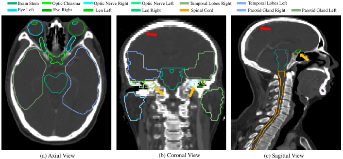
- 水晶体や視神経など、小さな、平坦で境界が曖昧なOARのセグメンテーション精度を向上させること。
- 放射線治療における臨床意思決定支援のための信頼性のある不確実性推定を提供すること。
- アノトロピックなCTスキャン(高スライス間隔)においてモデルの複雑さを低減しながら、性能を維持または向上させること。
- 強度変換、ハードボクセルへの注目、アンサンブルベースの不確実性評価を統合するフレームワークの構築
提案手法
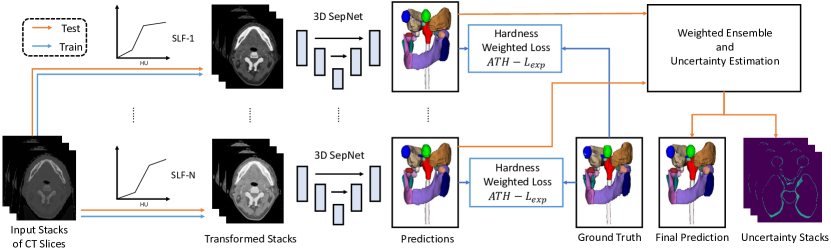
- 異なる組織タイプに応じて非一様な強度変換を適用することで、複数のOARを同時に可視化するためのセグメンテーション線形関数(SLF)を導入。
- 空間的分離可能3D畳み込みを用いた3D-SepNetアーキテクチャを提案し、高インプレーン解像度・低スライス間隔のアノトロピックCTスキャンを効率的に処理。
- ハードな領域に注目する損失関数(ATH-LExp)を設計。指数関数的損失と組み合わせることで、困難な領域の性能向上を実現。
- 異なるSLF強度変換および損失関数で訓練されたモデルをアンサンブル化することで、頑健なセグメンテーション出力と内在的な不確実性推定を生成。
- アンサンブル予測の分散を不確実性の代理指標として用い、誤分類領域と強く相関することが示された。
- 一般化を向上させ、不均衡なOARデータセットにおける過学習を低減するため、重み付き平均アンサンブル戦略を採用。
実験結果
リサーチクエスチョン
- RQ1標準的なウィンドウレベル調整と比較して、SLFのような学習可能な強度変換は、頭頸部CTにおけるマルチオルガニズム可視化およびセグメンテーション精度を向上させるか?
- RQ2高スライス間隔のアノトロピックCTスキャンからのOARセグメンテーションにおいて、標準的な3D UNetと比較して、分離可能3D畳み込みネットワーク(3D-SepNet)はどのように性能を発揮するか?
- RQ3ハードなボクセルに注目する損失関数は、特に小さなまたは低コントラストなOARにおいて、セグメンテーション精度を向上させるか?
- RQ4アンサンブルベースのモデル出力は、臨床現場におけるセグメンテーション誤差と相関する信頼性の高い不確実性推定を提供できるか?
- RQ5不確実性マップは、放射線治療計画における手動補正の対象とする高リスク領域に放射線科医を誘導するのにどの程度有効か?
主な発見
- 提案された3D-SepNetは、StructSeg 2019 HAN OARチャレンジにおいて、重み付き平均Diceスコア80.52%および95%ハウスドルフ距離3.043 mmを達成し、3位を獲得した。
- 3D UNetのパラメータ数の1/3で、特に視神経や水晶体のような平坦または薄い構造物において、セグメンテーション精度が優れていた。
- SLFに基づく強度変換は、複数のOARを同時に可視化することで、標準的な線形ウィンドウ処理を上回るセグメンテーション精度を著しく向上させた。
- ハードボクセルに注目するATH-LExp損失関数は、標準的なDSCや指数関数的損失単体よりも、特に小さなまたは境界が曖昧なOARにおいて高いDiceスコアを達成した。
- アンサンブル分散から導出された不確実性マップは、誤分類領域と強く相関しており、補正のための臨床的ガイダンスとしての強力な可能性を示した。
- 本手法は多様なデータセットに対して頑健であり、追加のトレーニングや推論コストを伴わず、実用的な不確実性情報の提供が可能であった。
より良い研究を、今すぐ始めましょう
論文の読解から最終レビューまで、研究時間を劇的に削減しましょう。
クレジットカード登録不要
このレビューはAIが作成し、人間の編集者が確認しました。