[論文レビュー] Breast Cancer Segmentation using Attention-based Convolutional Network and Explainable AI
この論文は、CLAHE 前処理と説明可能AI(Grad-CAM)を用いてモデルの焦点を分析した、超音波画像における乳がん分割のための注意機構を持つ UNet アーキテクチャを提案し、競合的な精度と IoU を達成します。
Breast cancer (BC) remains a significant health threat, with no long-term cure currently available. Early detection is crucial, yet mammography interpretation is hindered by high false positives and negatives. With BC incidence projected to surpass lung cancer, improving early detection methods is vital. Thermography, using high-resolution infrared cameras, offers promise, especially when combined with artificial intelligence (AI). This work presents an attention-based convolutional neural network for segmentation, providing increased speed and precision in BC detection and classification. The system enhances images and performs cancer segmentation with explainable AI. We propose a transformer-attention-based convolutional architecture (UNet) for fault identification and employ Gradient-weighted Class Activation Mapping (Grad-CAM) to analyze areas of bias and weakness in the UNet architecture with IRT images. The superiority of our proposed framework is confirmed when compared with existing deep learning frameworks.
研究の動機と目的
- マンモグラフィーと超音波画像診断における誤解を減らすための自動化された乳がん検出と分割の動機づけ。
- ROI 抽出を改善するために注意機構を持つ CNN を組み合わせたセグメンテーションフレームワークを開発。
- 超音波ベースの BC 分割におけるモデルの意思決定を可視化・理解するための説明可能AIを組み込む。
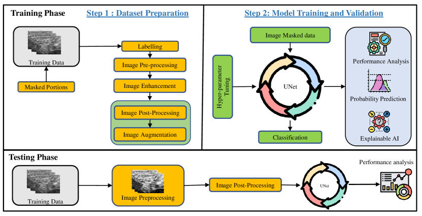
提案手法
- CLAHE によるヒストグラム均等化を用いて超音波画像を強化し、関心領域を抽出。
- Attention Gate を備えた UNet ベースのエンコーダ-デコーダアーキテクチャを採用し、分割品質を向上。
- 分割フレームワーク内に Vision Transformer(ViT)要素を組み込み、性能を強化。
- IoU、Loss、Accuracy、Precision、Recall、F1-Score を含む標準指標で分割を評価し、XAI 分析には Grad-CAM を使用。
- 提案手法の AttentionUNet を BC データセット上の最先端深層学習フレームワークと比較。
実験結果
リサーチクエスチョン
- RQ1注意機構を備えた UNet は標準アーキテクチャと比較して超音波画像の乳がん分割を改善できるか。
- RQ2CLAHE ベースの前処理はノイズの多い超音波データにおける ROI 抽出と分割性能を改善するか。
- RQ3Grad-CAM は AI ベースの分割に対する臨床的信頼を支える意味のある焦点領域の説明を提供できるか。
主な発見
- 最大のトレーニング精度は 95%、最大の検証精度は 99%。
- モデルは 12 エポック後に強い分割性能を示し、高い IoU と堅牢な学習曲線を示す。
- Grad-CAM 分析を用いてモデル領域を検査し、分割決定の説明可能性を支持する。
- 提案アーキテクチャは BC 超音波データセット上で他の既存の深層学習フレームワークよりも高速かつ精度が高いと主張される。
- 定性的な結果は、モデルが暗い領域を効果的に検出できる一方で不規則な形状には苦戦する可能性があることを示しており、トレーニングの調整(例: より長いトレーニング、分割エポック)を示唆している。

より良い研究を、今すぐ始めましょう
論文設計から論文執筆まで、研究時間を劇的に削減しましょう。
クレジットカード登録不要
このレビューはAIが作成し、人間の編集者が確認しました。