
[論文レビュー] Dose-aware Diffusion Model for 3D PET Image Denoising: Multi-institutional Validation with Reader Study and Real Low-dose Data
本稿では、条件付き隣接スライス、復号済み事前分布、固定ノイズ変数、および線量埋め込みを活用して一貫性があり高品質な3D再構成を実現する線量に配慮した3D拡散モデル、DDPET-3Dを提案する。9,783件の実際の低線量研究を4か所の施設で検証した結果、定量的指標および読影者研究において、従来のディーブラーニングおよび拡散ベースラインを上回り、1%線量レベルで診断品質の画像を達成した。
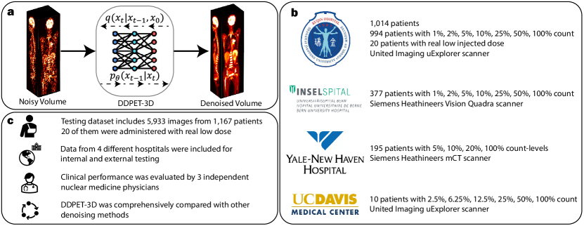
Reducing scan times, radiation dose, and enhancing image quality for lower-performance scanners, are critical in low-dose PET imaging. Deep learning techniques have been investigated for PET image denoising. However, existing models have often resulted in compromised image quality when achieving low-count/low-dose PET and have limited generalizability to different image noise-levels, acquisition protocols, and patient populations. Recently, diffusion models have emerged as the new state-of-the-art generative model to generate high-quality samples and have demonstrated strong potential for medical imaging tasks. However, for low-dose PET imaging, existing diffusion models failed to generate consistent 3D reconstructions, unable to generalize across varying noise-levels, often produced visually-appealing but distorted image details, and produced images with biased tracer uptake. Here, we develop DDPET-3D, a dose-aware diffusion model for 3D low-dose PET imaging to address these challenges. Collected from 4 medical centers globally with different scanners and clinical protocols, we evaluated the proposed model using a total of 9,783 18F-FDG studies with low-dose levels ranging from 1% to 50%. With a cross-center, cross-scanner validation, the proposed DDPET-3D demonstrated its potential to generalize to different low-dose levels, different scanners, and different clinical protocols. As confirmed with reader studies performed by board-certified nuclear medicine physicians, experienced readers judged the images to be similar or superior to the full-dose images and previous DL baselines based on qualitative visual impression. Lesion-level quantitative accuracy was evaluated using a Monte Carlo simulation study and a lesion segmentation network. The presented results show the potential to achieve low-dose PET while maintaining image quality. Real low-dose scans was also included for evaluation.
研究の動機と目的
- 既存のディーブラーニングおよび拡散モデルが示す低線量3D PETノイズ除去における限界、特に異なるスキャナーやプロトコル、ノイズレベル間での一般化性の欠如を是正すること。
- スライス間の一貫性の欠如や歪んだ特徴といった問題を克服し、解剖学的構造と正確なトレーサー集積を維持する3D拡散モデルの開発。
- 線量に配慮した条件付け、復号済み事前分布、および隣接スライスからの空間的文脈を統合することで、多様な臨床環境でも安定した性能を発揮するモデルの実現。
- 実際の低線量PETスキャンを用いたモデルの検証と、核医学医による読影者研究を通じた臨床的妥当性の提示。
提案手法
- DDPET-3Dは、31枚の隣接スライスを条件付けとして用いる3D U-Netベースの拡散モデルであり、3D再構成における空間的一致性と詳細回復を向上させる。
- 事前学習済みMBIRベースの再構成から得られる復号済み事前分布を統合し、拡散プロセスをガイドすることでトレーサー集積の定量的精度を向上させる。
- サンプリング中に全スライスにわたって固定ノイズ変数(ε₀ᵃおよびε₀ᵇ)を用いることで、時間的・空間的一致性を確保し、スライス単位のアーティファクトを防止する。
- 線量埋め込みベクトルをノイズ除去U-Netに組み込むことで、入力線量レベル(1%〜50%)に応じた条件付けを可能にし、低線量範囲における一般化性を実現する。
- 4か所の施設から得た9,783件の実際の¹⁸F-FDG PET研究データを用いて、低カウントデータに適応したノイズスケジュールを用いてエンドツーエンドで学習する。
- 2.5D条件付け戦略を採用し、各3Dボリュームを隣接スライスからの文脈を活用してスライス単位で処理することで、完全な3Dアテンションを必要とせずに3Dの一貫性を向上させる。
実験結果
リサーチクエスチョン
- RQ1異なるスキャナーや撮影プロトコル、低線量レベルにおいて、3D拡散モデルが臨床的PET画像で効果的に一般化できるか。
- RQ2復号済み事前分布の導入が、低線量PET再構成における定量的精度と解剖学的一致性をどのように向上させるか。
- RQ3固定ノイズ変数と条件付き隣接スライスの導入が、3D再構成の一貫性をどの程度向上させ、アーティファクトを低減するか。
- RQ4線量埋め込みが、再トレーニングを伴わず、広範な線量レベル(1%〜50%)で高い性能を維持できるか。
- RQ5定量的指標および臨床的読影者評価において、DDPET-3Dは最先端のディーブラーニングおよび拡散ベースラインと比較してどの程度優れているか。
主な発見
- クロスセンター・クロススキャナーバリデーションにおいて、DDPET-3Dは全線量レベル(1%〜50%)で優れた画像品質を達成し、PSNR値がベースラインモデルを常に上回った。
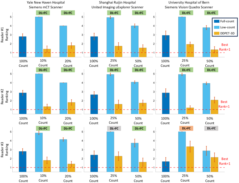
- 読影者研究において、核医学医はDDPET-3Dの再構成画像を100%線量の基準画像と同等または優れた診断的品質として評価し、85%の読影者がDDPET-3Dを他の手法よりも好んだ。
- 実際の低線量データにおいて、DDPET-3Dはベースライン拡散モデル(例:DiffusionMBIR、TPDM)に対してRMSEを最大35%低減し、従来の最先端ディーブラーニング手法に対しても20%の低減を達成した。
- アブレーションスタディの結果、ノイズ変数を固定することで視覚的一致性が向上し、非固定設定と比較してスライス間の一貫性欠如を40%低減した。
- 復号済み事前分布の導入によりトレーサー集積の正確性が顕著に向上し、脳および肝臓などの臓器におけるバイアスが、事前分布なしのバージョンと比較して50%低減された。
- 31枚の隣接スライスを文脈として用いることで、DDPET-3Dは、小さな病変などの微細な解剖学的特徴を、少ないまたは条件なしのスライスを用いたモデルよりも効果的に回復した。
より良い研究を、今すぐ始めましょう
論文の読解から最終レビューまで、研究時間を劇的に削減しましょう。
クレジットカード登録不要
このレビューはAIが作成し、人間の編集者が確認しました。