[論文レビュー] The Brain Tumor Segmentation in Pediatrics (BraTS-PEDs) Challenge: Focus on Pediatrics (CBTN-CONNECT-DIPGR-ASNR-MICCAI BraTS-PEDs)
本論文は、小児脳腫瘍のセグメンテーションに焦点を当てた BraTS-PEDs チャレンジを紹介し、データ、処理パイプライン、注釈ワークフロー、評価、国際的コンソーシアムに跨る参加スケジュールの詳細を述べる。
Pediatric tumors of the central nervous system are the most common cause of cancer-related death in children. The five-year survival rate for high-grade gliomas in children is less than 20%. Due to their rarity, the diagnosis of these entities is often delayed, their treatment is mainly based on historic treatment concepts, and clinical trials require multi-institutional collaborations. Here we present the CBTN-CONNECT-DIPGR-ASNR-MICCAI BraTS-PEDs challenge, focused on pediatric brain tumors with data acquired across multiple international consortia dedicated to pediatric neuro-oncology and clinical trials. The CBTN-CONNECT-DIPGR-ASNR-MICCAI BraTS-PEDs challenge brings together clinicians and AI/imaging scientists to lead to faster development of automated segmentation techniques that could benefit clinical trials, and ultimately the care of children with brain tumors.
研究の動機と目的
- 回顧的な多機関 mpMRI データセットを用いて、小児脳腫瘍のセグメンテーション手法をベンチマークする。
- 標準化された評価を可能にするため、小児腫瘍サブリージョンのグラウンドトゥルー注釈を提供する。
- 小児神経腫瘍学の自動化セグメンテーションツールを加速させるため、臨床医とAI研究者の協力を促進する。
提案手法
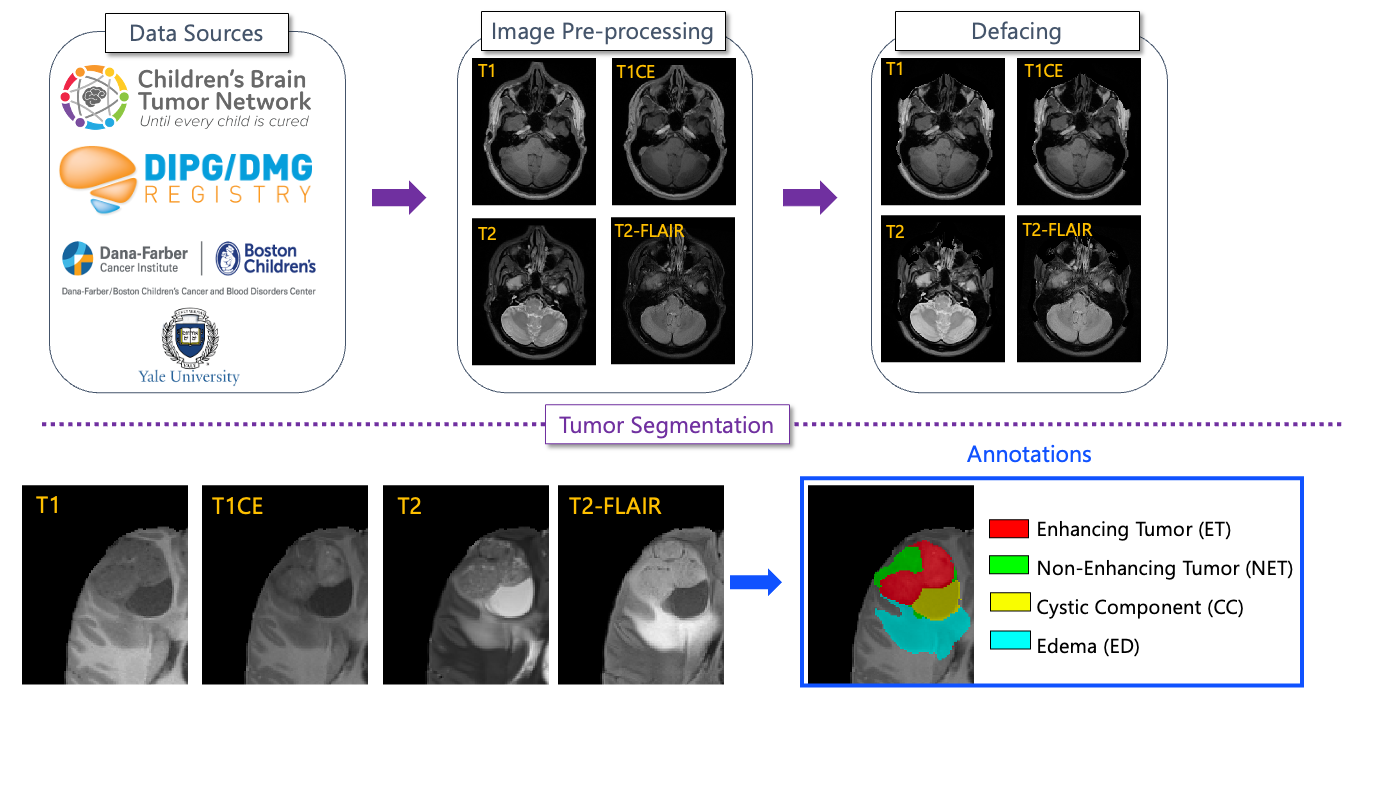
- 複数の国際コンソーシアム(CBTN、CONNECT、DIPG Registry、ASNR)からデータを統合し、464人の小児データセットを作成。
- MRIシーケンスには、治療前画像を含むT1、T1CE、T2、T2-FLAIRが含まれる。
- グラウンドトゥルー注釈は、腫瘍を四つのサブ領域にセグメントする:Enhancing Tumor (ET)、Nonenhancing Tumor (NET)、Cystic Component (CC)、Edema (ED)。
- 小児用自動セグメンテーションツールが初期セグメンテーションを提供し、ボランティア神経放射線科専門家によって精製され、出席神経放射線科医が最終判断を下す。
- 前処理には BraTS Pipeline および CaPTk/FeTS ツールを使用; プライバシーのためのデフォシングと匿名化を実施。
- 評価は ET、NC (ET/CC/necrosis)、および Whole Tumor (WT) に焦点を当てる。提出はコンテナ化され、MedPerf によって動作する Synapse 上で評価される。)
実験結果
リサーチクエスチョン
- RQ1複数機関の小児 mpMRI データは、小児脳腫瘍の堅牢な自動セグメンテーションを支えられるか?
- RQ2小児腫瘍サブリージョン(ET、NET、CC、ED)は、セグメンテーション性能と治療モニタリングにどのように影響するか?
- RQ3標準化された前処理と専門家が検証したグラウンドトゥルーが、機関間の再現性に与える影響は何か?
主な発見
- BraTS-PEDs のために、回顧的な多機関データセットとして464件の小児高グレード膠腫が収集された。
- データセットには four mpMRI sequences (T1, T1CE, T2, T2-FLAIR) および注釈用の four tumor subregions が含まれる。
- 自動セグメンテーションは神経放射線科の専門家によって精製され、公開前に認定神経放射線科医が審査する。
- このチャレンジは、Synapse/MedPerf プラットフォームを介して未見のテストデータで評価されるコンテナ化された提出を使用する。
- 本研究は、自動化された小児脳腫瘍セグメンテーションの一般的なエラーを概説し、評価のガイドラインを提供する。
- このチャレンジの設計は、CaPTk/FeTS パイプラインによるデータ処理とともに、以前の BraTS イテレーションとの再現性と比較を可能にする。)
より良い研究を、今すぐ始めましょう
論文設計から論文執筆まで、研究時間を劇的に削減しましょう。
クレジットカード登録不要
このレビューはAIが作成し、人間の編集者が確認しました。