[論文レビュー] Weakly supervised segmentation of intracranial aneurysms using a novel 3D focal modulation UNet
FocalSegNetを紹介する。TOF-MRA上の弱教師ありUIAセグメンテーションのための3D焦点モジュレーションUNetで、CRF後処理を用いた従来のUNetバリアントを上回る。
Accurate identification and quantification of unruptured intracranial aneurysms (UIAs) is crucial for the risk assessment and treatment of this cerebrovascular disorder. Current 2D manual assessment on 3D magnetic resonance angiography (MRA) is suboptimal and time-consuming. In addition, one major issue in medical image segmentation is the need for large well-annotated data, which can be expensive to obtain. Techniques that mitigate this requirement, such as weakly supervised learning with coarse labels are highly desirable. In the paper, we propose FocalSegNet, a novel 3D focal modulation UNet, to detect an aneurysm and offer an initial, coarse segmentation of it from time-of-flight MRA image patches, which is further refined with a dense conditional random field (CRF) post-processing layer to produce a final segmentation map. We trained and evaluated our model on a public dataset, and in terms of UIA detection, our model showed a low false-positive rate of 0.21 and a high sensitivity of 0.80. For voxel-wise aneurysm segmentation, we achieved a Dice score of 0.68 and a 95% Hausdorff distance of ~0.95 mm, demonstrating its strong performance. We evaluated our algorithms against the state-of-the-art 3D Residual-UNet and Swin-UNETR, and illustrated the superior performance of our proposed FocalSegNet, highlighting the advantages of employing focal modulation for this task.
研究の動機と目的
- 注釈付きデータが限られている状況で、TOF-MRAからのUIAセグメンテーションを高精度に行う動機づけ。
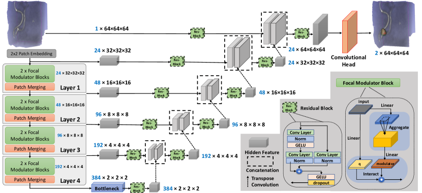
- 弱教師ありセグメンテーションのための新規3D焦点モジュレーション UNet(FocalSegNet)を提案します。
- 3D UIAセグメンテーションにおける焦点モジュレーションと自己注意の影響を調査する。
- 全結合CRF後処理ステップを用いてセグメンテーションを強化する。
- UIAセグメンテーション性能に影響を与える主要因を特定するためのアブレーション研究を提供する。
提案手法
- Swin-UnetRに似た UNetのエンコーダを3D焦点モジュレーションブロックと置換して3D FocalSegNetを開発する。
- TOF-MRAからの粗いUIAラベルで学習し、クラス不均衡に対処するために交差エントロピーと境界損失を用いたDice/IoUベースの損失を適用する。
- 初期予測を整えるために全結合CRFを後処理として適用する。
- 弱ラベリングの下でFocalSegNetを3D UNetおよびSwin-UNETRのベースラインと比較する。
- 解剖学的に情報を得たパッチ抽出とデータ増強を用いたパッチ単位のセグメーションを実行し、データのまれさを緩和する。
実験結果
リサーチクエスチョン
- RQ13D焦点モジュレーション UNetは、自己注意ベースのモデルと比較してTOF-MRA上のUIAの弱教師ありセグメンテーションを改善できるか?
- RQ2UIAのセグメンテーション精度と検出信頼性に対するCRF後処理の影響はどの程度か?
- RQ3高度に不均衡なUIAセグメンテーションにおいて、損失関数成分(交差エントロピー、一般化Dice、境界損失)は性能にどう影響するか?
- RQ43D医用画像セグメンテーションタスクにおいて、焦点モジュレーションは従来の自己注意より有利か?
主な発見
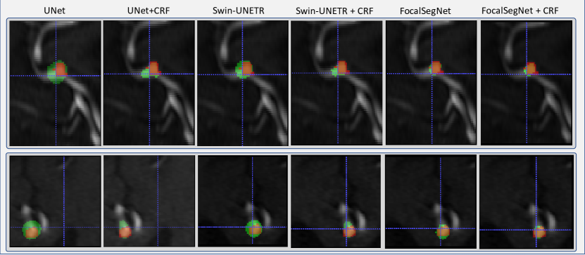
- CRFを用いたFocalSegNetはUIAセグメンテーションでDice 0.678±0.141、95-HD 2.148±1.082を達成し、UNetベースラインを上回る。
- FocalSegNetは偽陽性率を低く示し(0.212±0.464)、感度も競争力がある(0.801±0.399)、Swin-UNETRおよびUNet系と比較して。
- CRFなしでもFocalSegNetはすでに高い性能を示し、CRF後処理はモデル間で精度をさらに向上させる。
- Swin-UNETRと比較して、FocalSegNetは同等またはより良いセグメンテーション指標を示し、偽陽性率を有意に低下させる(FP比較でp<0.05)。
- アブレーションは、CRF後処理とCE、一般化Dice、および境界損失の組み合わせが最高性能に重要であることを示している。
より良い研究を、今すぐ始めましょう
論文設計から論文執筆まで、研究時間を劇的に削減しましょう。
クレジットカード登録不要
このレビューはAIが作成し、人間の編集者が確認しました。