
[Paper Review] Comment on newly found Charge Density Waves in infinite layer Nickelates
This paper challenges the attribution of a reported charge density wave (CDW) in infinite-layer nickelates (NdNiO₂/SrTiO₃) to resonant X-ray scattering, demonstrating that the signal arises from non-resonant contamination due to third-harmonic scattering from the SrTiO₃ substrate. Using fixed-momentum-transfer (E fixQ) scans, the authors show no resonance at the Ni L₃ edge, proving the signal is dominated by substrate Bragg peaks, and advocate E fixQ as the new standard for distinguishing true resonant responses in RIXS experiments.
Recent works[1-3] reported evidence for charge density waves (CDWs) in infinite layer nickelates (112 structure) based on resonant diffraction at the Ni $L_3$ edge measured at fixed scattering angle. We have found that a measurement with fixed momentum transfer, rather than scattering angle, does not show a resonance effect. We have also observed that a nearby structural Bragg peak from the substrate appears due to third harmonic content of the incident beam, and spreads intensity down to the region of the attributed CDW order. This was further confirmed by testing a bare substrate. We suggest procedures to confirm an effective resonant enhancement of a diffraction peak.
Motivation & Objective
- To critically assess the evidence for charge density waves (CDWs) in infinite-layer nickelates (NdNiO₂/SrTiO₃) reported via resonant inelastic X-ray scattering (RIXS).
- To investigate whether the observed elastic signal at Q_peak = (1/3, 0, 0.31) rlu is truly resonant or contaminated by non-resonant scattering from the SrTiO₃ substrate.
- To demonstrate that fixed-scattering-angle (E fix2θ) scans produce misleading resonant-like features due to momentum transfer drift, while fixed-momentum-transfer (E fixQ) scans reveal the true non-resonant nature of the signal.
- To propose E fixQ scanning as the new standard for identifying genuine resonant enhancements in quasi-elastic X-ray scattering, especially in 2D oxide heterostructures.
Proposed method
- Performed resonant elastic X-ray scattering (REXS) on NdNiO₂/SrTiO₃ and bare SrTiO₃ substrates at 100 K using the TARDIS endstation at NSLS-II.
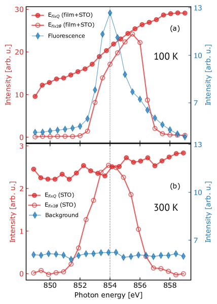
- Conducted two types of energy scans: E fix2θ (fixed scattering angle) and E fixQ (fixed momentum transfer), comparing their responses at Q_peak.
- Used background subtraction to isolate elastic scattering from fluorescence contributions, with data collected in σ-polarization geometry.
- Mapped the reciprocal lattice to identify proximity of Q_peak to the (1,0,1) Bragg peak of the SrTiO₃ substrate, which exhibits third-harmonic contamination.
- Measured the same Q_peak on a bare SrTiO₃ substrate to isolate substrate-specific scattering contributions.
- Calculated the substrate Bragg peak position as Q_STO = (1/3, 0, 0.28) rlu in film reciprocal lattice units, matching Q_peak within experimental uncertainty.
Experimental results
Research questions
- RQ1Is the reported CDW signal in NdNiO₂/SrTiO₃ truly resonant at the Ni L₃ edge, or is it an artifact of measurement geometry?
- RQ2To what extent does third-harmonic contamination from the SrTiO₃ substrate contribute to the observed elastic signal at Q_peak?
- RQ3Why do E fix2θ scans show a resonant-like peak while E fixQ scans show a continuously increasing signal with energy?
- RQ4Can the observed signal at Q_peak be fully explained by non-resonant scattering from the substrate rather than electronic CDW order?
- RQ5What experimental protocol ensures unambiguous identification of true resonant enhancements in RIXS and REXS measurements?
Key findings
- The E fix2θ scan shows a resonant-like peak centered at the Ni L₃ edge, consistent with prior RIXS reports, but this is an artifact of varying momentum transfer with energy.
- The E fixQ scan shows a continuously increasing signal across the Ni L₃ edge energy range with no peak, proving the dominant contribution is non-resonant.
- The elastic signal at Q_peak is primarily due to third-harmonic contamination from the SrTiO₃ substrate’s (1,0,1) Bragg peak, which has Q_STO = (1/3, 0, 0.28) rlu, matching Q_peak = (1/3, 0, 0.31) rlu within experimental uncertainty.
- On a bare SrTiO₃ substrate, E fix2θ still shows an apparent resonance at the Ni L₃ edge, while E fixQ shows constant intensity, confirming that the effect is substrate-driven and not electronic.
- The study demonstrates that E fixQ scanning is essential to distinguish true resonant responses from geometric and substrate-induced artifacts in X-ray scattering.
- The authors conclude that the reported CDW signal in NdNiO₂/SrTiO₃ is not reliably attributed to electronic order and propose E fixQ as the new standard for resonant scattering experiments.
Better researchstarts right now
From reading papers to final review, dramatically reduce your research time.
No credit card · Free plan available
This review was created by AI and reviewed by human editors.