
[Paper Review] FetMRQC: a robust quality control system for multi-centric fetal brain MRI
FetMRQC is an open-source, interpretable machine learning framework that automates quality control for multi-centric fetal brain MRI by fusing 332 image quality metrics into predictions using random forests. It achieves robust generalization across diverse scanners and protocols, outperforming baselines in both regression (R² = 0.78) and classification (F1 = 0.83) tasks on a large, multi-center dataset of 1,600+ manually rated T2-weighted fetal MR images.
Fetal brain MRI is becoming an increasingly relevant complement to neurosonography for perinatal diagnosis, allowing fundamental insights into fetal brain development throughout gestation. However, uncontrolled fetal motion and heterogeneity in acquisition protocols lead to data of variable quality, potentially biasing the outcome of subsequent studies. We present FetMRQC, an open-source machine-learning framework for automated image quality assessment and quality control that is robust to domain shifts induced by the heterogeneity of clinical data. FetMRQC extracts an ensemble of quality metrics from unprocessed anatomical MRI and combines them to predict experts' ratings using random forests. We validate our framework on a pioneeringly large and diverse dataset of more than 1600 manually rated fetal brain T2-weighted images from four clinical centers and 13 different scanners. Our study shows that FetMRQC's predictions generalize well to unseen data while being interpretable. FetMRQC is a step towards more robust fetal brain neuroimaging, which has the potential to shed new insights on the developing human brain.
Motivation & Objective
- Address the lack of standardized, automated quality control in multi-centric fetal brain MRI due to motion artifacts and protocol heterogeneity.
- Develop a framework that generalizes across diverse clinical scanners and acquisition protocols without requiring domain adaptation.
- Enable interpretable, automated assessment of image quality to support reliable data curation and downstream analysis in fetal neuroimaging.
- Reduce reliance on expert manual rating by providing a scalable, objective alternative for image quality assessment in large-scale studies.
Proposed method
- Extracts 332 image quality metrics (IQMs) from unprocessed T2-weighted fetal MRI, including intensity, structural, and deep learning-based features.
- Uses random forests to combine IQMs and predict expert ratings for image quality, ensuring robustness and interpretability.
- Employs a nested cross-validation strategy during initial model selection to avoid overfitting and optimize hyperparameters.
- Applies feature importance analysis to identify the 20 most predictive IQMs, enabling a lightweight variant (FetMRQC-20) with minimal performance loss.
- Validates performance using standard metrics: R², Spearman correlation, MAE for regression; F1-score, AUC, precision, and recall for classification.
- Integrates with super-resolution reconstruction pipelines to improve output quality by filtering out low-quality stacks.
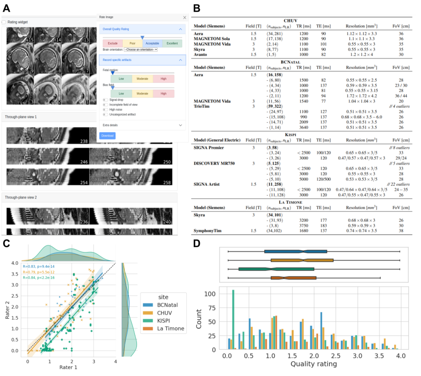
![Figure 1: Variations in data quality illustrated. A – Comparison of data across adult (T1w), from the ABIDE dataset [ 13 ] and from fetal acquisitions. In the excluded scans, the adult image on the left suffers from severe motion artifacts, while large coil artifacts corrupt the image on the right.](https://ar5iv.labs.arxiv.org/html/2311.04780/assets/x1.png)
Experimental results
Research questions
- RQ1Can a machine learning framework trained on multi-centric fetal MRI data generalize to unseen scanners and protocols without fine-tuning?
- RQ2Which combination of image quality metrics yields the most accurate and robust prediction of expert-rated image quality?
- RQ3How does FetMRQC compare to existing baselines, including deep learning and rule-based methods, in terms of predictive performance and generalization?
- RQ4To what extent does FetMRQC improve downstream processing, such as super-resolution reconstruction, by filtering low-quality data?
- RQ5Can the framework be made interpretable and transparent, allowing clinicians and researchers to understand the rationale behind quality predictions?
Key findings
- FetMRQC achieves a Pearson R² of 0.78 in regression and a weighted F1-score of 0.83 in classification, demonstrating strong predictive performance on unseen data.
- The framework generalizes effectively across four clinical centers and 13 different MRI scanners, showing robustness to domain shifts.
- The top 20 IQMs identified by feature importance (e.g., rank error, mask volume, normalized cross-correlation) account for most of the predictive power, enabling a lightweight variant (FetMRQC-20) with minimal performance drop.
- FetMRQC significantly improves super-resolution reconstruction quality by filtering out low-quality stacks, reducing artifacts and enhancing final image fidelity.
- The model outperforms established baselines, including NiftyMIC-QC and deep learning-based methods (dl_stack, dl_slice), especially in handling imbalanced quality distributions.
- Feature importance analysis reveals that structural metrics (e.g., mask volume, centroid) and motion-related features (e.g., rank error) are among the most influential predictors of image quality.
Better researchstarts right now
From reading papers to final review, dramatically reduce your research time.
No credit card · Free plan available
This review was created by AI and reviewed by human editors.