
[论文解读] Morphology-Enhanced CAM-Guided SAM for weakly supervised Breast Lesion Segmentation
该论文提出了一种形态学增强、CAM引导的SAM框架,用于仅使用图像级标注的超声图像乳腺病灶弱监督分割。通过融合病灶形态先验与基于类激活图(CAM)的定位,并利用分割一切模型(SAM)精炼预测,该方法在Dice分数上达到74.39%,在边界精度方面优于完全监督和弱监督基线方法。
Ultrasound imaging plays a critical role in the early detection of breast cancer. Accurate identification and segmentation of lesions are essential steps in clinical practice, requiring methods to assist physicians in lesion segmentation. However, ultrasound lesion segmentation models based on supervised learning require extensive manual labeling, which is both time-consuming and labor-intensive. In this study, we present a novel framework for weakly supervised lesion segmentation in early breast ultrasound images. Our method uses morphological enhancement and class activation map (CAM)-guided localization. Finally, we employ the Segment Anything Model (SAM), a computer vision foundation model, for detailed segmentation. This approach does not require pixel-level annotation, thereby reducing the cost of data annotation. The performance of our method is comparable to supervised learning methods that require manual annotations, achieving a Dice score of 74.39% and outperforming comparative supervised models in terms of Hausdorff distance in the BUSI dataset. These results demonstrate that our framework effectively integrates weakly supervised learning with SAM, providing a promising solution for breast cancer image analysis. The code for this study is available at: https://github.com/YueXin18/MorSeg-CAM-SAM.
研究动机与目标
- 减少在乳腺超声病灶分割中对昂贵像素级标注的依赖。
- 提升早期乳腺癌检测中不规则和小病灶的分割精度。
- 将医学先验知识(病灶形态)与基于深度学习的定位方法(CAM)及基础模型(SAM)相结合。
- 为资源有限的临床环境中的初级筛查开发一种可行且低资源的解决方案。
- 在边界精度和拓扑正确性方面超越现有的弱监督和完全监督方法。
提出的方法
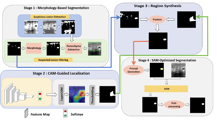
- 该方法首先通过形态学感知分割模块,利用解剖学先验提取病灶轮廓。
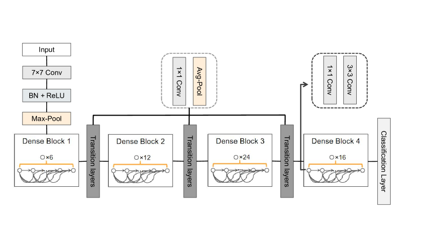
- 使用预训练的分类网络生成类激活图(CAM),基于语义特征定位病灶区域。
- 将形态学图与CAM融合,生成混合提示以引导分割一切模型(SAM)。
- SAM利用该融合提示执行精细化、高精度的分割,充分发挥其零样本泛化能力。
- 应用后处理以纠正拓扑错误,提升掩码的连通性与平滑度。
- 该框架仅使用图像级标签,无需像素级标注。
实验结果
研究问题
- RQ1将病灶形态先验与基于CAM的定位相结合,能否提升乳腺超声图像中弱监督分割的性能?
- RQ2形态学与CAM特征的融合如何增强基于SAM的分割性能?
- RQ3仅使用图像级标注的弱监督框架能否实现与完全监督模型相媲美的性能?
- RQ4整合医学先验知识是否能降低对昂贵像素级标注的依赖?
- RQ5与最先进的完全监督和弱监督模型相比,该方法在边界精度和病灶轮廓恢复方面表现如何?
主要发现
- 所提方法在BUSI测试集上达到74.39%的Dice分数,性能强劲,可与完全监督方法相媲美。
- 该方法在Hausdorff距离(HD95)上达到24.27,显著优于Deeplabv3+(32.22),表明边界定位能力更优。
- 该框架能有效分割小尺寸和不规则形状的病灶,在轮廓准确度方面优于UNet和Deeplabv3+。
- 消融实验表明,形态学、CAM和SAM各组件对最终性能均有显著贡献。
- 该方法通过过滤非病灶区域,有效减少了假阳性分割,提升了对背景噪声的鲁棒性。
- 该方法对噪声或模糊边界具有鲁棒性,后处理能有效纠正SAM引入的拓扑伪影。
更好的研究,从现在开始
从阅读论文到最终审阅,大幅缩短您的研究时间。
无需绑定信用卡
本解读由 AI 生成,并经人工编辑审核。